当前位置:
X-MOL 学术
›
Polym. Chem.
›
论文详情
Our official English website, www.x-mol.net, welcomes your
feedback! (Note: you will need to create a separate account there.)
An AIE-driven fluorescent polysaccharide polymersome as an enzyme-responsive FRET nanoprobe to study the real-time delivery aspects in live cells
Polymer Chemistry ( IF 4.1 ) Pub Date : 2020-12-18 , DOI: 10.1039/d0py01085e Nilesh Umakant Deshpande 1, 2, 3, 4 , Mishika Virmani 1, 2, 3, 4 , Manickam Jayakannan 1, 2, 3, 4
Polymer Chemistry ( IF 4.1 ) Pub Date : 2020-12-18 , DOI: 10.1039/d0py01085e Nilesh Umakant Deshpande 1, 2, 3, 4 , Mishika Virmani 1, 2, 3, 4 , Manickam Jayakannan 1, 2, 3, 4
Affiliation
|
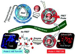
We report aggregation induced emission (AIE) driven polysaccharide polymersomes as fluorescence resonance energy transfer (FRET) nanoprobes to study their intracellular enzyme-responsive delivery by real-time live-cell confocal microscopy bio-imaging techniques. An AIE active tetraphenylethylene (TPE) optical chromophore and plant-based vesicular directing hydrophobic unit were grafted on clinically relevant polysaccharide-dextran via enzyme-cleavable aliphatic ester chemical linkages. The TPE-tagged dextran self-assembled as 180 ± 20 nm blue-luminescent polymersomes in aqueous medium and exhibited excellent encapsulation capabilities for water soluble Rose Bengal (RB) and water insoluble Nile red (NR) fluorophores. The selective photoexcitation of the TPE chromophore enabled the FRET process between the TPE donor and RB (or NR) acceptor molecule in <50 Å Förster distance afforded by the polymersome. The FRET probe was very stable under extracellular conditions and it exclusively underwent lysosomal esterase enzymatic biodegradation at the intracellular compartments to release RB. The enzyme-trigger enabled the FRET probe to function as an extracellular turn-ON → intracellular turn-Off red-fluorescent signal (Probe-1). In this process, the AIE self-emission was also simultaneously restored on the TPE chromophore (blue-luminescent, Probe-2) followed by the isolation of donor and acceptor in the cytosol. As a result, this new design enabled the visualization of real-time enzyme-responsive delivery by monitoring the dual fluorescent signals from both the polymer host (blue) and encapsulated guest (red) in a single nano-platform. In vitro cytotoxicity studies established that the polymersome probe was non-toxic to cells up to 300 μg mL−1. Lyso-tracker staining experiments supported the FRET probe internalization in the lysosomal compartments for enzymatic-biodegradation. Live cell confocal microscopy with selective photo-excitation was used to directly monitor the enzyme-responsive FRET action in human breast cancer MCF 7 and wild-type mouse embryonic fibroblast cell lines (WT-MEFs). It was found that the tailor-made polymersome FRET probe was efficient to deliver the loaded cargo in <3 h in live cells which predicts the usefulness of the probe in biomedical research.
中文翻译:

AIE驱动的荧光多糖多聚体作为酶反应性FRET纳米探针,用于研究活细胞中的实时递送方面
我们报告聚集诱导发射(AIE)驱动的多糖多聚体作为荧光共振能量转移(FRET)纳米探针,以通过实时活细胞共聚焦显微镜生物成像技术研究其细胞内酶反应性传递。AIE活性四苯基乙烯(TPE)光学发色团和植物基囊泡定向疏水单元通过以下方法嫁接到临床相关的多糖-葡聚糖上酶可裂解的脂族酯化学键。TPE标记的右旋糖酐在水性介质中自组装为180±20 nm的蓝色发光聚合物囊泡,对水溶性玫瑰红(RB)和水不溶性尼罗红(NR)荧光团表现出出色的封装能力。TPE生色团的选择性光激发使TPE供体与RB(或NR)受体分子之间的FRET过程在聚合体提供的<50ÅFörster距离内得以实现。FRET探针在细胞外条件下非常稳定,仅在细胞内区室进行溶酶体酯酶的酶促生物降解以释放RB。酶触发使FRET探针能够用作细胞外开启→细胞内关闭红色荧光信号(Probe-1)。在这个过程中 AIE自发光也同时在TPE生色团上恢复(蓝色发光,Probe-2),然后分离细胞质中的供体和受体。结果,该新设计通过监视来自单个纳米平台中聚合物主体(蓝色)和封装的客体(红色)的双重荧光信号,实现了实时酶响应递送的可视化。体外细胞毒性研究确定,多聚体探针对高达300μgmL -1的细胞无毒。溶菌跟踪染色实验支持溶酶体区室中的FRET探针内化,以进行酶促生物降解。具有选择性光激发的活细胞共聚焦显微镜用于直接监测人乳腺癌MCF 7和野生型小鼠胚胎成纤维细胞系(WT-MEF)中的酶响应FRET作用。结果发现,量身定制的多聚体FRET探针可有效地在不到3小时的时间内将负载的货物递送到活细胞中,这预示了该探针在生物医学研究中的有用性。
更新日期:2021-01-12
中文翻译:
AIE驱动的荧光多糖多聚体作为酶反应性FRET纳米探针,用于研究活细胞中的实时递送方面
我们报告聚集诱导发射(AIE)驱动的多糖多聚体作为荧光共振能量转移(FRET)纳米探针,以通过实时活细胞共聚焦显微镜生物成像技术研究其细胞内酶反应性传递。AIE活性四苯基乙烯(TPE)光学发色团和植物基囊泡定向疏水单元通过以下方法嫁接到临床相关的多糖-葡聚糖上酶可裂解的脂族酯化学键。TPE标记的右旋糖酐在水性介质中自组装为180±20 nm的蓝色发光聚合物囊泡,对水溶性玫瑰红(RB)和水不溶性尼罗红(NR)荧光团表现出出色的封装能力。TPE生色团的选择性光激发使TPE供体与RB(或NR)受体分子之间的FRET过程在聚合体提供的<50ÅFörster距离内得以实现。FRET探针在细胞外条件下非常稳定,仅在细胞内区室进行溶酶体酯酶的酶促生物降解以释放RB。酶触发使FRET探针能够用作细胞外开启→细胞内关闭红色荧光信号(Probe-1)。在这个过程中 AIE自发光也同时在TPE生色团上恢复(蓝色发光,Probe-2),然后分离细胞质中的供体和受体。结果,该新设计通过监视来自单个纳米平台中聚合物主体(蓝色)和封装的客体(红色)的双重荧光信号,实现了实时酶响应递送的可视化。体外细胞毒性研究确定,多聚体探针对高达300μgmL -1的细胞无毒。溶菌跟踪染色实验支持溶酶体区室中的FRET探针内化,以进行酶促生物降解。具有选择性光激发的活细胞共聚焦显微镜用于直接监测人乳腺癌MCF 7和野生型小鼠胚胎成纤维细胞系(WT-MEF)中的酶响应FRET作用。结果发现,量身定制的多聚体FRET探针可有效地在不到3小时的时间内将负载的货物递送到活细胞中,这预示了该探针在生物医学研究中的有用性。


 京公网安备 11010802027423号