当前位置:
X-MOL 学术
›
NMR Biomed.
›
论文详情
Our official English website, www.x-mol.net, welcomes your
feedback! (Note: you will need to create a separate account there.)
The muscle twitch profile assessed with motor unit magnetic resonance imaging
NMR in Biomedicine ( IF 2.7 ) Pub Date : 2021-01-06 , DOI: 10.1002/nbm.4466 Linda Heskamp 1 , Matthew G Birkbeck 1, 2, 3 , Roger G Whittaker 1 , Ian S Schofield 1 , Andrew M Blamire 1
NMR in Biomedicine ( IF 2.7 ) Pub Date : 2021-01-06 , DOI: 10.1002/nbm.4466 Linda Heskamp 1 , Matthew G Birkbeck 1, 2, 3 , Roger G Whittaker 1 , Ian S Schofield 1 , Andrew M Blamire 1
Affiliation
|
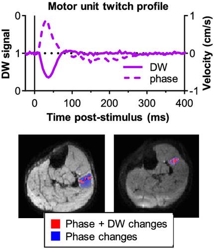
Localised signal voids in diffusion‐weighted (DW) images of skeletal muscle have been postulated to occur as a result of muscle fibre contraction and relaxation. We investigated the contrast mechanism of these signal voids using a combination of modelling and experimental measurements by employing DW and phase contrast (PC) imaging sequences. The DW signal and PC signal were simulated for each time point of a theoretical muscle twitch. The model incorporated compaction (simulating actively contracting muscle fibres) and translation (simulating passively moving surrounding fibres). The model suggested that the DW signal depended on contraction time and compaction whereas the PC signal depended on contraction time, compaction and translation. In a retrospective study, we tested this model with subgroup analyses on 10 healthy participants. Electrical nerve stimulation was used to generate muscle twitches in lower leg muscles; the resulting force was measured using an MR‐compatible force transducer. At current levels causing a visible muscle twitch (~13 mA), the width of the first signal drop in the DW signal (mean ± SD: 103 ± 20 ms) was comparable with the force contraction time (93 ± 34 ms; intraclass correlation coefficient [ICC] = 0.717, P = .010). At current levels activating single motor units (~9 mA), the contraction time determined from the DW signal was 75 ± 13 ms and comparable with the PC contraction time (81 ± 15 ms; ICC = 0.925, P = .001). The maximum positive velocity was 0.55 ± 0.26 cm/s and the displacement was 0.20 ± 0.10 mm. Voxel‐wise analysis revealed localised DW changes occurring together with more widespread phase changes. In conclusion, local signal attenuations in DW images following muscle fibre activation are primarily caused by compaction. The PC sequence also detects translating muscle tissue being passively pulled. The magnitude of the changes in DW and PC images depends on the twitch's contractile properties and percentage contraction. DW imaging and PC imaging can therefore measure twitch profiles of skeletal muscle fibres.
中文翻译:

用运动单元磁共振成像评估的肌肉抽搐特征
骨骼肌的弥散加权 (DW) 图像中的局部信号空白被假定为肌肉纤维收缩和松弛的结果。我们通过采用 DW 和相位对比 (PC) 成像序列,结合建模和实验测量研究了这些信号空隙的对比机制。DW 信号和 PC 信号针对理论肌肉抽搐的每个时间点进行模拟。该模型结合了压实(模拟主动收缩的肌肉纤维)和平移(模拟被动移动周围的纤维)。该模型表明 DW 信号取决于收缩时间和压实,而 PC 信号取决于收缩时间、压实和平移。在一项回顾性研究中,我们通过对 10 名健康参与者的亚组分析测试了该模型。电神经刺激用于在小腿肌肉中产生肌肉抽搐;使用 MR 兼容的力传感器测量产生的力。在导致可见肌肉抽搐 (~13 mA) 的当前水平下,DW 信号中第一个信号下降的宽度(平均值 ± SD:103 ± 20 毫秒)与力收缩时间(93 ± 34 毫秒;组内相关性)相当系数 [ICC] = 0.717,P = .010)。在激活单个运动单元 (~9 mA) 的当前水平下,根据 DW 信号确定的收缩时间为 75 ± 13 ms,与 PC 收缩时间相当(81 ± 15 ms;ICC = 0.925,P= .001)。最大正速度为 0.55 ± 0.26 cm/s,位移为 0.20 ± 0.10 mm。体素分析显示局部 DW 变化与更广泛的相位变化一起发生。总之,肌肉纤维激活后 DW 图像中的局部信号衰减主要是由压实引起的。PC 序列还检测被被动拉动的平移肌肉组织。DW 和 PC 图像的变化幅度取决于抽搐的收缩特性和收缩百分比。因此,DW 成像和 PC 成像可以测量骨骼肌纤维的抽搐剖面。
更新日期:2021-02-04
中文翻译:
用运动单元磁共振成像评估的肌肉抽搐特征
骨骼肌的弥散加权 (DW) 图像中的局部信号空白被假定为肌肉纤维收缩和松弛的结果。我们通过采用 DW 和相位对比 (PC) 成像序列,结合建模和实验测量研究了这些信号空隙的对比机制。DW 信号和 PC 信号针对理论肌肉抽搐的每个时间点进行模拟。该模型结合了压实(模拟主动收缩的肌肉纤维)和平移(模拟被动移动周围的纤维)。该模型表明 DW 信号取决于收缩时间和压实,而 PC 信号取决于收缩时间、压实和平移。在一项回顾性研究中,我们通过对 10 名健康参与者的亚组分析测试了该模型。电神经刺激用于在小腿肌肉中产生肌肉抽搐;使用 MR 兼容的力传感器测量产生的力。在导致可见肌肉抽搐 (~13 mA) 的当前水平下,DW 信号中第一个信号下降的宽度(平均值 ± SD:103 ± 20 毫秒)与力收缩时间(93 ± 34 毫秒;组内相关性)相当系数 [ICC] = 0.717,P = .010)。在激活单个运动单元 (~9 mA) 的当前水平下,根据 DW 信号确定的收缩时间为 75 ± 13 ms,与 PC 收缩时间相当(81 ± 15 ms;ICC = 0.925,P= .001)。最大正速度为 0.55 ± 0.26 cm/s,位移为 0.20 ± 0.10 mm。体素分析显示局部 DW 变化与更广泛的相位变化一起发生。总之,肌肉纤维激活后 DW 图像中的局部信号衰减主要是由压实引起的。PC 序列还检测被被动拉动的平移肌肉组织。DW 和 PC 图像的变化幅度取决于抽搐的收缩特性和收缩百分比。因此,DW 成像和 PC 成像可以测量骨骼肌纤维的抽搐剖面。


 京公网安备 11010802027423号