当前位置:
X-MOL 学术
›
Microsc. Res. Tech.
›
论文详情
Our official English website, www.x-mol.net, welcomes your
feedback! (Note: you will need to create a separate account there.)
Filling quality of several obturation techniques in the presence of apically separated instruments: A Micro-CT study
Microscopy Research and Technique ( IF 2.0 ) Pub Date : 2020-12-30 , DOI: 10.1002/jemt.23685 Sevinç Aktemur Türker 1 , Emel Uzunoğlu-Özyürek 2 , Sena Kaşikçi 1 , Melike Öndeş 3 , Ferhat Geneci 4 , Hakan Hamdi Çelik 5
Microscopy Research and Technique ( IF 2.0 ) Pub Date : 2020-12-30 , DOI: 10.1002/jemt.23685 Sevinç Aktemur Türker 1 , Emel Uzunoğlu-Özyürek 2 , Sena Kaşikçi 1 , Melike Öndeş 3 , Ferhat Geneci 4 , Hakan Hamdi Çelik 5
Affiliation
|
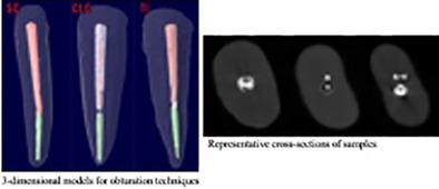
Separated root canal instruments may affect the quality of root canal obturation and hence the survival of endodontically treated teeth. Obturation techniques may influence filling-quality. The aim was to evaluate the obturation quality of teeth filled with different obturation techniques in the presence of apically separated instruments using micro-computed tomography (μ-CT). Notched ProTaper F2 rotary-files were separated in the apical third of 36-human mandibular incisors with single root/canal and mature apex. Samples were filled by an endodontist according to one of the following obturation techniques (n = 12): cold lateral-compaction (CLC), single-cone (SC) and thermoplastic injection (TI). Samples were scanned by the Skyscan 1,274® μ-CT device after 1-week at 37°C in 100% humidity. Images of the sections were evaluated with CTan software in terms of total-volume and volume percentage of the filling materials and voids between coronal end of the separated instrument and gutta-percha/sealer filled void-free sections. Data was analyzed using Kruskal-Wallis and Mann Whitney-U tests with a significance level of 5%. Kruskal–Wallis revealed differences among groups (p < 0.05).Pairwise comparisons revealed that less volume of voids were measured in SC (0.02 ± 0.03 mm3) compared to CLC (0.15 ± 0.16 mm3) and TI (0.18 ± 0.24 mm3) (p < 0.05); while TI was statistically similar with CLC (p > 0.05). Percentages of volumes of voids and filling materials were as follows for SC, CLC and TI, respectively: 8.88 ± 18.52% and 24.45 ± 38.40%, 46.92 ± 33.53% and 53.07 ± 33.53%, 40.54 ± 33.85% and 42.79 ± 34.45%. The obturation technique may have a significant impact on the volume of voids in the presence of a separated file. No obturation technique produced a void-free root canal filling.
中文翻译:

在存在顶部分离器械的情况下几种充填技术的填充质量:微 CT 研究
分开的根管器械可能会影响根管封闭的质量,从而影响经过牙髓治疗的牙齿的存活率。填充技术可能会影响填充质量。目的是使用微型计算机断层扫描 (μ-CT) 评估在存在根尖分离器械的情况下用不同充填技术填充的牙齿的充填质量。在具有单根/根管和成熟根尖的 36 人下颌切牙的根尖三分之一处分离有缺口的 ProTaper F2 旋转锉。样本由牙髓病医生根据以下填充技术之一进行填充 ( n = 12):冷横向压实 (CLC)、单锥 (SC) 和热塑性塑料注射 (TI)。在 37°C 和 100% 湿度下,1 周后,使用 Skyscan 1,274® μ-CT 设备扫描样品。使用 CTan 软件根据填充材料的总体积和体积百分比以及分离器械的冠状端与牙胶/密封剂填充的无空隙部分之间的空隙对切片的图像进行评估。使用 Kruskal-Wallis 和 Mann Whitney-U 检验分析数据,显着性水平为 5%。Kruskal-Wallis 揭示了组间差异 ( p < 0.05)。成对比较显示,与 CLC (0.15 ± 0.16 mm 3 ) 和 TI (0.18 ± 0.24 mm 3 ) 相比,SC (0.02 ± 0.03 mm 3 )中测量到的空隙体积更小) (p < 0.05);而 TI 与 CLC 在统计学上相似(p > 0.05)。SC、CLC 和 TI 的空隙和填充材料的体积百分比分别如下:8.88 ± 18.52% 和 24.45 ± 38.40%、46.92 ± 33.53% 和 53.07 ± 33.53%、40.54 ± 33.24.7% 和 34.34.85% 在分离锉存在的情况下,密闭技术可能对空隙的体积有显着影响。没有充填技术产生无空隙的根管充填。
更新日期:2020-12-30
中文翻译:
在存在顶部分离器械的情况下几种充填技术的填充质量:微 CT 研究
分开的根管器械可能会影响根管封闭的质量,从而影响经过牙髓治疗的牙齿的存活率。填充技术可能会影响填充质量。目的是使用微型计算机断层扫描 (μ-CT) 评估在存在根尖分离器械的情况下用不同充填技术填充的牙齿的充填质量。在具有单根/根管和成熟根尖的 36 人下颌切牙的根尖三分之一处分离有缺口的 ProTaper F2 旋转锉。样本由牙髓病医生根据以下填充技术之一进行填充 ( n = 12):冷横向压实 (CLC)、单锥 (SC) 和热塑性塑料注射 (TI)。在 37°C 和 100% 湿度下,1 周后,使用 Skyscan 1,274® μ-CT 设备扫描样品。使用 CTan 软件根据填充材料的总体积和体积百分比以及分离器械的冠状端与牙胶/密封剂填充的无空隙部分之间的空隙对切片的图像进行评估。使用 Kruskal-Wallis 和 Mann Whitney-U 检验分析数据,显着性水平为 5%。Kruskal-Wallis 揭示了组间差异 ( p < 0.05)。成对比较显示,与 CLC (0.15 ± 0.16 mm 3 ) 和 TI (0.18 ± 0.24 mm 3 ) 相比,SC (0.02 ± 0.03 mm 3 )中测量到的空隙体积更小) (p < 0.05);而 TI 与 CLC 在统计学上相似(p > 0.05)。SC、CLC 和 TI 的空隙和填充材料的体积百分比分别如下:8.88 ± 18.52% 和 24.45 ± 38.40%、46.92 ± 33.53% 和 53.07 ± 33.53%、40.54 ± 33.24.7% 和 34.34.85% 在分离锉存在的情况下,密闭技术可能对空隙的体积有显着影响。没有充填技术产生无空隙的根管充填。


 京公网安备 11010802027423号