Spectrochimica Acta Part B: Atomic Spectroscopy ( IF 3.2 ) Pub Date : 2020-12-26 , DOI: 10.1016/j.sab.2020.106063 Buddhadev Kanrar , Kaushik Sanyal , Sangita Dhara
|
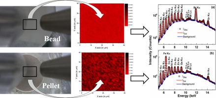
Pressed pelletization and fusion bead methods are two very important sample preparation techniques for X-Ray Fluorescence (XRF) analysis. Hence, along with the assessment of these two techniques with respect to its analytical capabilities, it is also very important to study the elemental homogeneity in the prepared specimens. The distribution of trace elements in the specimen prepared using pressed pelletization and fusion bead method were studied using a recently installed table top Micro-XRF (μ-XRF) spectrometer. The spectrometer is equipped with focussing polycapillary lens to obtain the X-ray spot size of 51.3 μm × 33.5 μm. An area of 4 mm × 4 mm was scanned using μ-XRF and the data obtained were used for the elemental mapping of the trace elements in those sample specimens. It was observed that the distribution of trace elements in the specimens depend on their concentration. The elements were distributed more homogeneously in bead than in pellet specimens and the homogeneity increases with the elemental concentration in both the specimens. The μ-XRF determined elemental concentrations of the trace elements in fused bead and pressed pellet specimens were in good agreement with the expected values. However for lower elemental concentration, it was observed that fused bead specimen technique is better. The fused bead technique was utilised to determine trace elemental concentration in a real soil sample.
中文翻译:
使用台式微X射线荧光光谱仪对熔融珠和压丸样品中的微量元素进行定量和分布
压丸法和熔融珠法是用于X射线荧光(XRF)分析的两种非常重要的样品制备技术。因此,连同对这两种技术的分析能力进行评估一样,研究制备样品中的元素均质性也非常重要。使用最近安装的台式Micro-XRF(μ-XRF)光谱仪研究了使用压丸法和熔融珠法制备的样品中痕量元素的分布。光谱仪配备了聚焦多毛细管透镜,以获得51.3μm×33.5μm的X射线光斑尺寸。使用μ-XRF扫描4 mm×4 mm的面积,并将获得的数据用于这些样品中痕量元素的元素映射。据观察,样品中微量元素的分布取决于其浓度。元素在颗粒中的分布比在颗粒样品中更均匀,并且同质性随两个样品中元素浓度的增加而增加。μ-XRF测定的熔融珠和压丸样品中痕量元素的元素浓度与预期值非常吻合。但是,对于较低的元素浓度,可以观察到熔融珠试样技术更好。熔融珠技术用于确定真实土壤样品中的微量元素浓度。μ-XRF测定的熔融珠和压丸样品中痕量元素的元素浓度与预期值非常吻合。但是,对于较低的元素浓度,可以观察到熔融珠试样技术更好。熔融珠技术用于确定真实土壤样品中的微量元素浓度。μ-XRF测定的熔融珠和压丸样品中痕量元素的元素浓度与预期值非常吻合。但是,对于较低的元素浓度,可以观察到熔融珠试样技术更好。熔融珠技术用于确定真实土壤样品中的微量元素浓度。


 京公网安备 11010802027423号