当前位置:
X-MOL 学术
›
J. Morphol.
›
论文详情
Our official English website, www.x-mol.net, welcomes your
feedback! (Note: you will need to create a separate account there.)
Microvascular anatomy of the urinary bladder in the adult African clawed toad, Xenopus laevis : A scanning electron microscope study of vascular casts
Journal of Morphology ( IF 1.5 ) Pub Date : 2020-12-25 , DOI: 10.1002/jmor.21310 Alois Lametschwandtner 1 , Bernd Minnich 1
Journal of Morphology ( IF 1.5 ) Pub Date : 2020-12-25 , DOI: 10.1002/jmor.21310 Alois Lametschwandtner 1 , Bernd Minnich 1
Affiliation
|
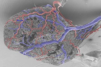
We studied urinary bladders of adult male and female Xenopus laevis using light microscopy of stained tissue sections and scanning electron microscopy (SEM) of vascular corrosion casts (VCCs). Results showed that bilaterally a vesical artery branched off the femoral artery. At the dorso-lateral serosal surface of the body of the bladder each artery splitted within a short distance into up to five smaller arteries that supplied body and neck regions. Arteries gave off short and long terminal arterioles, which fed the mucosal capillary meshwork. Long terminal arterioles followed dimensional changes of the bladder, while short ones anchored the capillary network to the arterial system. Capillary mesh sizes and shapes varied according to the filling state of the urinary bladder. In the highly to moderately distended (filled) bladder, capillaries were rather straight or undulated only slightly, in the contracted (emptied) bladder they undulated strongly and lay side by side. Postcapillary venules formed by two equally sized capillaries or from capillaries, which serially drained into a small postcapillary venule. Vesical venules formed a large dorsal vesical and a varying number of smaller lateral and ventral vesical veins. The dorsal vesical vein drained either directly or via the posterior hemorrhoidal vein into the common pelvic vein. Lateral and ventral vesical veins also drained into the latter. The vascular patterns found were discussed in respect to the bladder spatial movements during distention (filling) and relaxation (emptying). Furthermore, it was hypothesized that an extensively filled bladder could compress the overlaying abdominal vein forcing part of the blood otherwise drained towards the liver to be detoured via the renal portal veins to the kidneys.
中文翻译:

成年非洲爪蟾(非洲爪蟾)膀胱的微血管解剖:血管管型的扫描电子显微镜研究
我们使用染色组织切片的光学显微镜和血管腐蚀铸件 (VCC) 的扫描电子显微镜 (SEM) 研究了成年雄性和雌性非洲爪蟾的膀胱。结果显示双侧膀胱动脉从股动脉分支。在膀胱体的背外侧浆膜表面,每条动脉在短距离内分裂成多达五个较小的动脉,供应身体和颈部区域。动脉发出短小动脉和长末端小动脉,它们供给粘膜毛细血管网。长末端小动脉跟随膀胱的尺寸变化,而短末端小动脉将毛细血管网络锚定到动脉系统。毛细血管网的大小和形状根据膀胱的充盈状态而变化。在高度至中度膨胀(充满)的膀胱中,毛细血管相当直或仅轻微波动,在收缩(排空)的膀胱中,它们强烈波动并并排放置。毛细血管后微静脉由两条大小相等的毛细血管或毛细血管形成,依次汇入一个小的毛细血管后微静脉。膀胱小静脉形成了一个大的背侧膀胱和数量不等的较小的侧静脉和腹侧膀胱静脉。膀胱背静脉直接或经痔后静脉引流至盆腔总静脉。外侧和腹侧膀胱静脉也流入后者。在膨胀(充盈)和松弛(排空)期间,就膀胱空间运动对发现的血管模式进行了讨论。此外,
更新日期:2020-12-25
中文翻译:
成年非洲爪蟾(非洲爪蟾)膀胱的微血管解剖:血管管型的扫描电子显微镜研究
我们使用染色组织切片的光学显微镜和血管腐蚀铸件 (VCC) 的扫描电子显微镜 (SEM) 研究了成年雄性和雌性非洲爪蟾的膀胱。结果显示双侧膀胱动脉从股动脉分支。在膀胱体的背外侧浆膜表面,每条动脉在短距离内分裂成多达五个较小的动脉,供应身体和颈部区域。动脉发出短小动脉和长末端小动脉,它们供给粘膜毛细血管网。长末端小动脉跟随膀胱的尺寸变化,而短末端小动脉将毛细血管网络锚定到动脉系统。毛细血管网的大小和形状根据膀胱的充盈状态而变化。在高度至中度膨胀(充满)的膀胱中,毛细血管相当直或仅轻微波动,在收缩(排空)的膀胱中,它们强烈波动并并排放置。毛细血管后微静脉由两条大小相等的毛细血管或毛细血管形成,依次汇入一个小的毛细血管后微静脉。膀胱小静脉形成了一个大的背侧膀胱和数量不等的较小的侧静脉和腹侧膀胱静脉。膀胱背静脉直接或经痔后静脉引流至盆腔总静脉。外侧和腹侧膀胱静脉也流入后者。在膨胀(充盈)和松弛(排空)期间,就膀胱空间运动对发现的血管模式进行了讨论。此外,


 京公网安备 11010802027423号