当前位置:
X-MOL 学术
›
J. Neurosci. Res.
›
论文详情
Our official English website, www.x-mol.net, welcomes your feedback! (Note: you will need to create a separate account there.)
Detection of functional connectivity in the brain during visuo‐guided grip force tracking tasks: A functional near‐infrared spectroscopy study
Journal of Neuroscience Research ( IF 4.2 ) Pub Date : 2020-12-23 , DOI: 10.1002/jnr.24769 Xinyi Zheng 1 , Jie Luo 1 , Lingyun Deng 1 , Bing Li 1 , Le Li 2, 3 , Dong Feng Huang 3, 4 , Rong Song 1
Journal of Neuroscience Research ( IF 4.2 ) Pub Date : 2020-12-23 , DOI: 10.1002/jnr.24769 Xinyi Zheng 1 , Jie Luo 1 , Lingyun Deng 1 , Bing Li 1 , Le Li 2, 3 , Dong Feng Huang 3, 4 , Rong Song 1
Affiliation
|
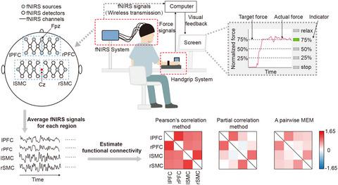
The functional connectivity (FC) between multiple brain regions during tasks is currently gradually being explored with functional near‐infrared spectroscopy (fNIRS). However, the FC present during grip force tracking tasks performed under visual feedback remains unclear. In the present study, we used fNIRS to measure brain activity during resting states and grip force tracking tasks at 25%, 50%, and 75% of maximum voluntary contraction (MVC) in 11 healthy subjects, and the activity was measured from four target brain regions: the left prefrontal cortex (lPFC), right prefrontal cortex (rPFC), left sensorimotor cortex (lSMC), and right sensorimotor cortex (rSMC). We determined the FC between these regions utilizing three different methods: Pearson's correlation method, partial correlation method, and a pairwise maximum entropy model (MEM). The results showed that the FC of lSMC‐rSMC and lPFC‐rPFC (interhemispheric homologous pairs) were significantly stronger than those of other brain region pairs. Moreover, FC of lPFC‐rPFC was strengthened during the 75% MVC task compared to the other task states and the resting states. The FC of lSMC‐lPFC and rSMC‐rPFC (intrahemispheric region pairs) strengthened with a higher task load. The results provided new insights into the FC between brain regions during visuo‐guided grip force tracking tasks.
中文翻译:

在视觉引导的握力跟踪任务中检测大脑中的功能连接:一项功能性近红外光谱研究
目前正在通过功能性近红外光谱 (fNIRS) 逐步探索任务期间多个大脑区域之间的功能连接性 (FC)。然而,在视觉反馈下执行的握力跟踪任务期间存在的 FC 仍不清楚。在本研究中,我们使用 fNIRS 测量了 11 名健康受试者在静息状态下的大脑活动和最大自主收缩 (MVC) 的 25%、50% 和 75% 的握力跟踪任务,并从四个目标测量了活动大脑区域:左前额叶皮层 (lPFC)、右前额叶皮层 (rPFC)、左感觉运动皮层 (lSMC) 和右感觉运动皮层 (rSMC)。我们使用三种不同的方法确定这些区域之间的 FC:Pearson 相关方法、偏相关方法和成对最大熵模型 (MEM)。结果表明,lSMC-rSMC 和 lPFC-rPFC(半球间同源对)的 FC 显着强于其他脑区对。此外,与其他任务状态和静止状态相比,lPFC-rPFC 的 FC 在 75% MVC 任务期间得到加强。lSMC-lPFC 和 rSMC-rPFC(半球内区域对)的 FC 因更高的任务负荷而增强。该结果为视觉引导握力跟踪任务期间大脑区域之间的 FC 提供了新的见解。lSMC-lPFC 和 rSMC-rPFC(半球内区域对)的 FC 因更高的任务负荷而增强。该结果为视觉引导握力跟踪任务期间大脑区域之间的 FC 提供了新的见解。lSMC-lPFC 和 rSMC-rPFC(半球内区域对)的 FC 因更高的任务负荷而增强。该结果为视觉引导握力跟踪任务期间大脑区域之间的 FC 提供了新的见解。
更新日期:2021-02-21
中文翻译:
在视觉引导的握力跟踪任务中检测大脑中的功能连接:一项功能性近红外光谱研究
目前正在通过功能性近红外光谱 (fNIRS) 逐步探索任务期间多个大脑区域之间的功能连接性 (FC)。然而,在视觉反馈下执行的握力跟踪任务期间存在的 FC 仍不清楚。在本研究中,我们使用 fNIRS 测量了 11 名健康受试者在静息状态下的大脑活动和最大自主收缩 (MVC) 的 25%、50% 和 75% 的握力跟踪任务,并从四个目标测量了活动大脑区域:左前额叶皮层 (lPFC)、右前额叶皮层 (rPFC)、左感觉运动皮层 (lSMC) 和右感觉运动皮层 (rSMC)。我们使用三种不同的方法确定这些区域之间的 FC:Pearson 相关方法、偏相关方法和成对最大熵模型 (MEM)。结果表明,lSMC-rSMC 和 lPFC-rPFC(半球间同源对)的 FC 显着强于其他脑区对。此外,与其他任务状态和静止状态相比,lPFC-rPFC 的 FC 在 75% MVC 任务期间得到加强。lSMC-lPFC 和 rSMC-rPFC(半球内区域对)的 FC 因更高的任务负荷而增强。该结果为视觉引导握力跟踪任务期间大脑区域之间的 FC 提供了新的见解。lSMC-lPFC 和 rSMC-rPFC(半球内区域对)的 FC 因更高的任务负荷而增强。该结果为视觉引导握力跟踪任务期间大脑区域之间的 FC 提供了新的见解。lSMC-lPFC 和 rSMC-rPFC(半球内区域对)的 FC 因更高的任务负荷而增强。该结果为视觉引导握力跟踪任务期间大脑区域之间的 FC 提供了新的见解。


 京公网安备 11010802027423号