当前位置:
X-MOL 学术
›
NMR Biomed.
›
论文详情
Our official English website, www.x-mol.net, welcomes your
feedback! (Note: you will need to create a separate account there.)
Improved preclinical hyperpolarized 129Xe ventilation imaging with constant flip angle 3D radial golden means acquisition and keyhole reconstruction
NMR in Biomedicine ( IF 2.7 ) Pub Date : 2020-12-23 , DOI: 10.1002/nbm.4464 Peter J Niedbalski 1, 2 , Zackary I Cleveland 1, 3, 4, 5
NMR in Biomedicine ( IF 2.7 ) Pub Date : 2020-12-23 , DOI: 10.1002/nbm.4464 Peter J Niedbalski 1, 2 , Zackary I Cleveland 1, 3, 4, 5
Affiliation
|
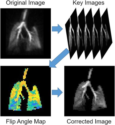
Hyperpolarized (HP) 129Xe MRI is increasingly used to noninvasively probe regional lung structure and function in the preclinical setting. As in human imaging, the primary barrier to quantitative imaging with HP gases is nonequilibrium magnetization, which is depleted by T1 relaxation and radio frequency excitation. Preclinical HP gas imaging commonly involves mechanically ventilating small animals and encoding k‐space over tens or hundreds of breaths, with small subsets of k‐space data collected within each breath. Breath‐to‐breath magnetization renewal enables the use of large flip angles, but the resulting magnetization decay generates large view‐to‐view differences in within‐breath signal intensity, leading to artifacts and degraded image quality. This deleterious signal decay has motivated the use of variable flip angle (VFA) sampling schemes, in which the flip angle is progressively increased to maintain constant view‐to‐view signal intensity. However, VFA imaging complicates data acquisition and provides only a global correction that fails to compensate for regional differences in signal dynamics. When constant flip angle (CFA) imaging is used alongside 3D radial golden means acquisition, the center of k‐space is sampled with every excitation, thereby encoding signal dynamics alongside imaging data. Here, keyhole reconstruction is used to generate multiple images to capture in‐breath HP 129Xe signal dynamics in mice and thus provide flip angle maps to quantitatively correct images without extra data collection. These CFA images display SNR that is not significantly different from VFA images, and further, high frequency k‐space scaling can be used to mitigate decay‐induced image artifacts. Results are supported by point spread function calculations and simulations of radial imaging with preclinical signal dynamics. Together, these results show that CFA 3D radial golden means ventilation imaging provides comparable image quality with VFA in small animals and allows for keyhole reconstruction, which can be used to generate flip angle maps and correct images for signal depletion.
中文翻译:

改进的临床前超极化 129Xe 通气成像,具有恒定翻转角 3D 径向黄金均值采集和锁孔重建
超极化 (HP) 129 Xe MRI 越来越多地用于在临床前环境中无创地探测局部肺结构和功能。与人类成像一样,HP 气体定量成像的主要障碍是非平衡磁化,它会被 T 1弛豫和射频激励耗尽。临床前 HP 气体成像通常涉及对小动物进行机械通气,并对数十或数百次呼吸的 k 空间进行编码,并在每次呼吸中收集 k 空间数据的小子集。呼吸间磁化更新可以使用大翻转角,但由此产生的磁化衰减会在呼吸内信号强度中产生较大的视图间差异,从而导致伪影和图像质量下降。这种有害的信号衰减促使人们使用可变翻转角(VFA)采样方案,其中翻转角逐渐增加以保持恒定的视图到视图信号强度。然而,VFA 成像使数据采集变得复杂,并且仅提供全局校正,无法补偿信号动态的区域差异。当恒定翻转角 (CFA) 成像与 3D 径向黄金均值采集一起使用时,每次激励都会对 k 空间中心进行采样,从而对信号动态与成像数据进行编码。在这里,锁孔重建用于生成多个图像以捕获小鼠的呼吸 HP 129 Xe 信号动态,从而提供翻转角度图以定量校正图像,而无需额外的数据收集。这些 CFA 图像显示的 SNR 与 VFA 图像没有显着差异,此外,高频 k 空间缩放可用于减轻衰变引起的图像伪影。 结果得到点扩散函数计算和具有临床前信号动力学的径向成像模拟的支持。总之,这些结果表明,CFA 3D 径向黄金均值通气成像可在小动物中提供与 VFA 相当的图像质量,并允许锁孔重建,可用于生成翻转角图并校正信号损耗的图像。
更新日期:2021-02-04
中文翻译:
改进的临床前超极化 129Xe 通气成像,具有恒定翻转角 3D 径向黄金均值采集和锁孔重建
超极化 (HP) 129 Xe MRI 越来越多地用于在临床前环境中无创地探测局部肺结构和功能。与人类成像一样,HP 气体定量成像的主要障碍是非平衡磁化,它会被 T 1弛豫和射频激励耗尽。临床前 HP 气体成像通常涉及对小动物进行机械通气,并对数十或数百次呼吸的 k 空间进行编码,并在每次呼吸中收集 k 空间数据的小子集。呼吸间磁化更新可以使用大翻转角,但由此产生的磁化衰减会在呼吸内信号强度中产生较大的视图间差异,从而导致伪影和图像质量下降。这种有害的信号衰减促使人们使用可变翻转角(VFA)采样方案,其中翻转角逐渐增加以保持恒定的视图到视图信号强度。然而,VFA 成像使数据采集变得复杂,并且仅提供全局校正,无法补偿信号动态的区域差异。当恒定翻转角 (CFA) 成像与 3D 径向黄金均值采集一起使用时,每次激励都会对 k 空间中心进行采样,从而对信号动态与成像数据进行编码。在这里,锁孔重建用于生成多个图像以捕获小鼠的呼吸 HP 129 Xe 信号动态,从而提供翻转角度图以定量校正图像,而无需额外的数据收集。这些 CFA 图像显示的 SNR 与 VFA 图像没有显着差异,此外,高频 k 空间缩放可用于减轻衰变引起的图像伪影。 结果得到点扩散函数计算和具有临床前信号动力学的径向成像模拟的支持。总之,这些结果表明,CFA 3D 径向黄金均值通气成像可在小动物中提供与 VFA 相当的图像质量,并允许锁孔重建,可用于生成翻转角图并校正信号损耗的图像。


 京公网安备 11010802027423号