当前位置:
X-MOL 学术
›
NMR Biomed.
›
论文详情
Our official English website, www.x-mol.net, welcomes your
feedback! (Note: you will need to create a separate account there.)
A high‐throughput imaging platform to characterize extracellular pH in organotypic three‐dimensional in vitro models of liver cancer
NMR in Biomedicine ( IF 2.7 ) Pub Date : 2020-12-22 , DOI: 10.1002/nbm.4465 Lynn Jeanette Savic 1, 2, 3 , Isabel Theresa Schobert 1, 2 , Charlie Alexander Hamm 1, 2, 4 , Lucas Christoph Adam 1, 2 , Fahmeed Hyder 1, 5 , Daniel Coman 1
NMR in Biomedicine ( IF 2.7 ) Pub Date : 2020-12-22 , DOI: 10.1002/nbm.4465 Lynn Jeanette Savic 1, 2, 3 , Isabel Theresa Schobert 1, 2 , Charlie Alexander Hamm 1, 2, 4 , Lucas Christoph Adam 1, 2 , Fahmeed Hyder 1, 5 , Daniel Coman 1
Affiliation
|
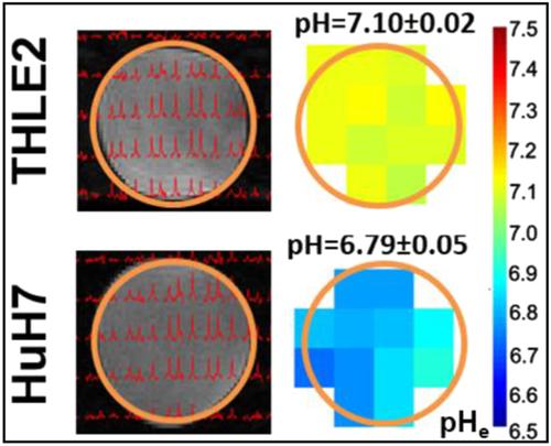
Given the extraordinary nature of tumor metabolism in hepatocellular carcinoma and its impact on oncologic treatment response, this study introduces a novel high‐throughput extracellular pH (pHe) mapping platform using magnetic resonance spectroscopic imaging in a three‐dimensional (3D) in vitro model of liver cancer. pHe mapping was performed using biosensor imaging of redundant deviation in shifts (BIRDS) on 9.4 T and 11.7 T MR scanners for validation purposes. 3D cultures of four liver cancer (HepG2, Huh7, SNU475, VX2) and one hepatocyte (THLE2) cell line were simultaneously analyzed (a) without treatment, (b) supplemented with 4.5 g/L d‐glucose, and (c) treated with anti‐glycolytic 3‐bromopyruvate (6.25, 25, 50, 75, and 100 μM). The MR results were correlated with immunohistochemistry (GLUT‐1, LAMP‐2) and luminescence‐based viability assays. Statistics included the unpaired t‐test and ANOVA test. High‐throughput pHe imaging with BIRDS for in vitro 3D liver cancer models proved feasible. Compared with non‐tumorous hepatocytes (pHe = 7.1 ± 0.1), acidic pHe was revealed in liver cancer (VX2, pHe = 6.7 ± 0.1; HuH7, pHe = 6.8 ± 0.1; HepG2, pHe = 6.9 ± 0.1; SNU475, pHe = 6.9 ± 0.1), in agreement with GLUT‐1 upregulation. Glucose addition significantly further decreased pHe in hyperglycolytic cell lines (VX2, HepG2, and Huh7, by 0.28, 0.06, and 0.11, respectively, all p < 0.001), whereas 3‐bromopyruvate normalized tumor pHe in a dose‐dependent manner without affecting viability. In summary, this study introduces a non‐invasive pHe imaging platform for high‐yield screening using a translational 3D liver cancer model, which may help reveal and target mechanisms of therapy resistance and inform personalized treatment of patients with hepatocellular carcinoma.
中文翻译:

用于表征肝癌器官型三维体外模型中细胞外 pH 值的高通量成像平台
鉴于肝细胞癌中肿瘤代谢的特殊性质及其对肿瘤治疗反应的影响,本研究在三维(3D)体外模型中使用磁共振光谱成像引入了一种新的高通量细胞外 pH(pH e)映射平台肝癌。pH e映射是在 9.4 T 和 11.7 T MR 扫描仪上使用偏移冗余偏差 (BIRDS) 的生物传感器成像进行的,以进行验证。同时分析了四种肝癌(HepG2、Huh7、SNU475、VX2)和一种肝细胞(THLE2)细胞系的 3D 培养物(a)未经处理,(b)补充 4.5 g/L d-葡萄糖,和 (c) 用抗糖酵解 3-溴丙酮酸(6.25、25、50、75 和 100 μM)处理。MR 结果与免疫组织化学(GLUT-1、LAMP-2)和基于发光的活力测定相关。统计包括非配对t检验和方差分析。BIRDS 用于体外 3D 肝癌模型的高通量 pH e成像被证明是可行的。与非肿瘤肝细胞(pH e = 7.1 ± 0.1)相比,肝癌(VX2,pH e = 6.7 ± 0.1;HuH7,pH e = 6.8 ± 0.1;HepG2,pH e = 6.9 ± 0.1 )显示酸性pH e ; SNU475, pH e= 6.9 ± 0.1),与 GLUT-1 上调一致。添加葡萄糖显著进一步降低pH值ê在hyperglycolytic细胞系(VX2,HepG2细胞,和Huh7,0.28,0.06,和0.11分别所有p <0.001),而3-溴丙酮酸盐标准化肿瘤pH值ê以剂量依赖的方式,而不影响生存能力。总之,本研究介绍了一种使用平移 3D 肝癌模型进行高产筛查的非侵入性 pH e成像平台,这可能有助于揭示和靶向治疗抵抗的机制,并为肝细胞癌患者的个性化治疗提供信息。
更新日期:2021-02-04
中文翻译:
用于表征肝癌器官型三维体外模型中细胞外 pH 值的高通量成像平台
鉴于肝细胞癌中肿瘤代谢的特殊性质及其对肿瘤治疗反应的影响,本研究在三维(3D)体外模型中使用磁共振光谱成像引入了一种新的高通量细胞外 pH(pH e)映射平台肝癌。pH e映射是在 9.4 T 和 11.7 T MR 扫描仪上使用偏移冗余偏差 (BIRDS) 的生物传感器成像进行的,以进行验证。同时分析了四种肝癌(HepG2、Huh7、SNU475、VX2)和一种肝细胞(THLE2)细胞系的 3D 培养物(a)未经处理,(b)补充 4.5 g/L d-葡萄糖,和 (c) 用抗糖酵解 3-溴丙酮酸(6.25、25、50、75 和 100 μM)处理。MR 结果与免疫组织化学(GLUT-1、LAMP-2)和基于发光的活力测定相关。统计包括非配对t检验和方差分析。BIRDS 用于体外 3D 肝癌模型的高通量 pH e成像被证明是可行的。与非肿瘤肝细胞(pH e = 7.1 ± 0.1)相比,肝癌(VX2,pH e = 6.7 ± 0.1;HuH7,pH e = 6.8 ± 0.1;HepG2,pH e = 6.9 ± 0.1 )显示酸性pH e ; SNU475, pH e= 6.9 ± 0.1),与 GLUT-1 上调一致。添加葡萄糖显著进一步降低pH值ê在hyperglycolytic细胞系(VX2,HepG2细胞,和Huh7,0.28,0.06,和0.11分别所有p <0.001),而3-溴丙酮酸盐标准化肿瘤pH值ê以剂量依赖的方式,而不影响生存能力。总之,本研究介绍了一种使用平移 3D 肝癌模型进行高产筛查的非侵入性 pH e成像平台,这可能有助于揭示和靶向治疗抵抗的机制,并为肝细胞癌患者的个性化治疗提供信息。


 京公网安备 11010802027423号