当前位置:
X-MOL 学术
›
Microsc. Res. Tech.
›
论文详情
Our official English website, www.x-mol.net, welcomes your
feedback! (Note: you will need to create a separate account there.)
Classification of retinal images based on convolutional neural network
Microscopy Research and Technique ( IF 2.0 ) Pub Date : 2020-12-22 , DOI: 10.1002/jemt.23596 Noha A El-Hag 1 , Ahmed Sedik 2 , Walid El-Shafai 3 , Heba M El-Hoseny 4 , Ashraf A M Khalaf 1 , Adel S El-Fishawy 2 , Waleed Al-Nuaimy 5 , Fathi E Abd El-Samie 3, 6 , Ghada M El-Banby 7
Microscopy Research and Technique ( IF 2.0 ) Pub Date : 2020-12-22 , DOI: 10.1002/jemt.23596 Noha A El-Hag 1 , Ahmed Sedik 2 , Walid El-Shafai 3 , Heba M El-Hoseny 4 , Ashraf A M Khalaf 1 , Adel S El-Fishawy 2 , Waleed Al-Nuaimy 5 , Fathi E Abd El-Samie 3, 6 , Ghada M El-Banby 7
Affiliation
|
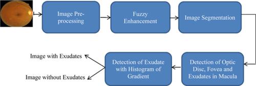
Automatic detection of maculopathy disease is a very important step to achieve high‐accuracy results for the early discovery of the disease to help ophthalmologists to treat patients. Manual detection of diabetic maculopathy needs much effort and time from ophthalmologists. Detection of exudates from retinal images is applied for the maculopathy disease diagnosis. The first proposed framework in this paper for retinal image classification begins with fuzzy preprocessing in order to improve the original image to enhance the contrast between the objects and the background. After that, image segmentation is performed through binarization of the image to extract both blood vessels and the optic disc and then remove them from the original image. A gradient process is performed on the retinal image after this removal process for discrimination between normal and abnormal cases. Histogram of the gradients is estimated, and consequently the cumulative histogram of gradients is obtained and compared with a threshold cumulative histogram at certain bins. To determine the threshold cumulative histogram, cumulative histograms of images with exudates and images without exudates are obtained and averaged for each type, and the threshold cumulative histogram is set as the average of both cumulative histograms. Certain histogram bins are selected and thresholded according to the estimated threshold cumulative histogram, and the results are used for retinal image classification. In the second framework in this paper, a Convolutional Neural Network (CNN) is utilized to classify normal and abnormal cases.
中文翻译:

基于卷积神经网络的视网膜图像分类
黄斑病变疾病的自动检测是获得高精度结果的非常重要的一步,可以及早发现疾病,帮助眼科医生治疗患者。手动检测糖尿病性黄斑病变需要眼科医生付出很多努力和时间。从视网膜图像中检测渗出物可用于黄斑病变疾病的诊断。本文提出的第一个视网膜图像分类框架从模糊预处理开始,以改进原始图像以增强物体与背景之间的对比度。之后,通过图像的二值化进行图像分割,提取血管和视盘,然后从原始图像中去除。在这个去除过程之后对视网膜图像执行梯度过程以区分正常和异常情况。估计梯度的直方图,从而获得梯度的累积直方图,并将其与某些区间的阈值累积直方图进行比较。为确定阈值累积直方图,获取有渗出物图像和无渗出物图像的累积直方图,并针对每种类型取平均值,并将阈值累积直方图设置为两种累积直方图的平均值。根据估计的阈值累积直方图选择某些直方图块并对其进行阈值处理,并将结果用于视网膜图像分类。在本文的第二个框架中,
更新日期:2021-02-15
中文翻译:
基于卷积神经网络的视网膜图像分类
黄斑病变疾病的自动检测是获得高精度结果的非常重要的一步,可以及早发现疾病,帮助眼科医生治疗患者。手动检测糖尿病性黄斑病变需要眼科医生付出很多努力和时间。从视网膜图像中检测渗出物可用于黄斑病变疾病的诊断。本文提出的第一个视网膜图像分类框架从模糊预处理开始,以改进原始图像以增强物体与背景之间的对比度。之后,通过图像的二值化进行图像分割,提取血管和视盘,然后从原始图像中去除。在这个去除过程之后对视网膜图像执行梯度过程以区分正常和异常情况。估计梯度的直方图,从而获得梯度的累积直方图,并将其与某些区间的阈值累积直方图进行比较。为确定阈值累积直方图,获取有渗出物图像和无渗出物图像的累积直方图,并针对每种类型取平均值,并将阈值累积直方图设置为两种累积直方图的平均值。根据估计的阈值累积直方图选择某些直方图块并对其进行阈值处理,并将结果用于视网膜图像分类。在本文的第二个框架中,


 京公网安备 11010802027423号