当前位置:
X-MOL 学术
›
Am. J. Primatol.
›
论文详情
Our official English website, www.x-mol.net, welcomes your
feedback! (Note: you will need to create a separate account there.)
The character of parietal and orbital alterations in the superfamily Hominoidea (families Hominidae [exclusive of Homo] and Hylobotidae)
American Journal of Primatology ( IF 2.0 ) Pub Date : 2020-12-21 , DOI: 10.1002/ajp.23227 Bruce Rothschild 1
American Journal of Primatology ( IF 2.0 ) Pub Date : 2020-12-21 , DOI: 10.1002/ajp.23227 Bruce Rothschild 1
Affiliation
|
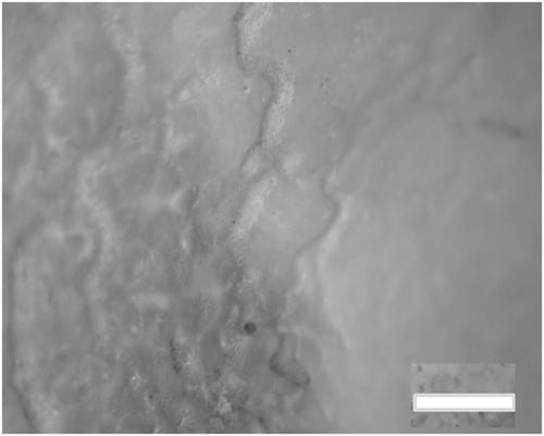
Parietal external surface disruption routinely referred to as porotic hyperostosis, and orbital alterations (cribra orbitalia), have been attributed to anemia‐related bone marrow hyperplasia in humans. A recent study in humans identified that they were actually vascular in nature. Skeletons were examined and epi‐illumination surface microscopy was performed on the parietal region and orbit of 156 Hominidae and 123 Hylobotidae to assess if these phenomena were trans‐phylogenetic. Trans‐cortical channels were recognized on the basis of visualized ectocranial surface defects penetrating the parietal; cribra orbitalia, by alteration of the normally smooth orbital roof appearance. Trans‐cortical parietal channels, ranging in size from 20 to 100 µm, are rare in Gorilla and Pan troglodytes and absent in Pan paniscus. They are universally present in adult Pongo abeli and in Hylobatidae, independent of species. Cribra orbitalia was common in Hylobotidae, Pongo pygmaeus and P. abelii, less prevalent in adult P. troglodytes, and not recognized in any Gorilla gorilla or P. paniscus examined. The proliferative form predominated, with the exception of Hylobates concolor and muelleri, in which uncalcified vascular grooves predominated. No correlation was observed between the presence of either trans‐cortical channels or cribra orbitalia and fractures, osteoarthritis, or inflammatory arthritis. Parietal alterations observed in apes are trans‐cortical channels, analogous to those observed in humans, and do not represent porosity. Similarly, cribra orbitalia in apes is confirmed as vascular in nature. The proliferative form apparently represents calcification of blood vessel walls, indistinguishable from observations in humans. Predominant presence in adults rather than in juveniles suggests that both forms are acquired rather than developmental in derivation. Sex and bone alteration/disease‐independence suggests that mechanical, endocrine, and inflammatory phenomena do not contribute to the development of either. Further, independent occurrence of trans‐cortical channels and cribra orbitalia suggests that they do not have a shared etiology.
中文翻译:

超人科(人科(不包括人)和Hylobotidae)的顶叶和眼眶改变的特征
壁外表面破坏通常被称为多孔性骨质增生,而眼眶的改变(cribra orbitalia)被归因于人类贫血相关的骨髓增生。一项针对人体的最新研究表明,它们实际上是血管。检查了骨骼并在顶叶区域和156个Homoboteidae和123个Hylobotidae的顶壁区域和轨道上进行了落射照明表面显微镜检查,以评估这些现象是否是跨系统发生的。通过可视化的颅骨表面缺陷穿透顶叶,可以识别出皮质通道。通过改变通常光滑的轨道屋顶外观,来改善cribra轨道。皮质顶壁通道的大小从20到100 µm不等,在大猩猩和潘中很少见。潘氏藻中的穴居动物和缺席。它们普遍存在于成年Pongo abeli和Hylobatidae中,而与物种无关。在Hylobotidae,Pongo pygmaeus和P. abelii中,斑马眼眶很常见,在成年P. troglodytes中较不普遍,并且在所检查的任何大猩猩大猩猩或P. paniscus中均未发现。增生形式占主导地位,但杂色的Hylobates和muelleri除外,其中未钙化的血管沟占主导。经皮皮质通道或斑马眼眶和骨折,骨关节炎或炎症性关节炎之间没有相关性。在猿类中观察到的顶壁改变是经皮层通道,类似于在人类中观察到的通道,并不代表孔隙。同样,猿猴中的天秤座轨道自然也被证实为血管。增生形式显然代表血管壁的钙化,与人类的观察结果没有区别。在成年人中而不是在青少年中占主导地位表明这两种形式都是后天获得而不是发育。性别和骨骼改变/疾病独立性表明,机械,内分泌和炎症现象均不会促进二者的发展。进一步,
更新日期:2021-01-04
中文翻译:
超人科(人科(不包括人)和Hylobotidae)的顶叶和眼眶改变的特征
壁外表面破坏通常被称为多孔性骨质增生,而眼眶的改变(cribra orbitalia)被归因于人类贫血相关的骨髓增生。一项针对人体的最新研究表明,它们实际上是血管。检查了骨骼并在顶叶区域和156个Homoboteidae和123个Hylobotidae的顶壁区域和轨道上进行了落射照明表面显微镜检查,以评估这些现象是否是跨系统发生的。通过可视化的颅骨表面缺陷穿透顶叶,可以识别出皮质通道。通过改变通常光滑的轨道屋顶外观,来改善cribra轨道。皮质顶壁通道的大小从20到100 µm不等,在大猩猩和潘中很少见。潘氏藻中的穴居动物和缺席。它们普遍存在于成年Pongo abeli和Hylobatidae中,而与物种无关。在Hylobotidae,Pongo pygmaeus和P. abelii中,斑马眼眶很常见,在成年P. troglodytes中较不普遍,并且在所检查的任何大猩猩大猩猩或P. paniscus中均未发现。增生形式占主导地位,但杂色的Hylobates和muelleri除外,其中未钙化的血管沟占主导。经皮皮质通道或斑马眼眶和骨折,骨关节炎或炎症性关节炎之间没有相关性。在猿类中观察到的顶壁改变是经皮层通道,类似于在人类中观察到的通道,并不代表孔隙。同样,猿猴中的天秤座轨道自然也被证实为血管。增生形式显然代表血管壁的钙化,与人类的观察结果没有区别。在成年人中而不是在青少年中占主导地位表明这两种形式都是后天获得而不是发育。性别和骨骼改变/疾病独立性表明,机械,内分泌和炎症现象均不会促进二者的发展。进一步,


 京公网安备 11010802027423号