当前位置:
X-MOL 学术
›
Clin. Exp. Immunol.
›
论文详情
Our official English website, www.x-mol.net, welcomes your
feedback! (Note: you will need to create a separate account there.)
Reconnaissance of tumor immune microenvironment spatial heterogeneity in metastatic renal cell carcinoma and correlation with immunotherapy response
Clinical & Experimental Immunology ( IF 3.4 ) Pub Date : 2020-12-21 , DOI: 10.1111/cei.13567 A Hajiran 1 , N Chakiryan 1 , A M Aydin 1 , L Zemp 1 , J Nguyen 2 , J M Laborde 3 , J Chahoud 1 , P E Spiess 1 , S Zaman 1 , S Falasiri 1 , M Fournier 1 , J K Teer 3 , J Dhillon 2 , S McCarthy 2 , C Moran-Segura 2 , E N Katende 1 , W J Sexton 1 , J M Koomen 4 , J Mulé 5 , Y Kim 2 , B Manley 1, 6
Clinical & Experimental Immunology ( IF 3.4 ) Pub Date : 2020-12-21 , DOI: 10.1111/cei.13567 A Hajiran 1 , N Chakiryan 1 , A M Aydin 1 , L Zemp 1 , J Nguyen 2 , J M Laborde 3 , J Chahoud 1 , P E Spiess 1 , S Zaman 1 , S Falasiri 1 , M Fournier 1 , J K Teer 3 , J Dhillon 2 , S McCarthy 2 , C Moran-Segura 2 , E N Katende 1 , W J Sexton 1 , J M Koomen 4 , J Mulé 5 , Y Kim 2 , B Manley 1, 6
Affiliation
|
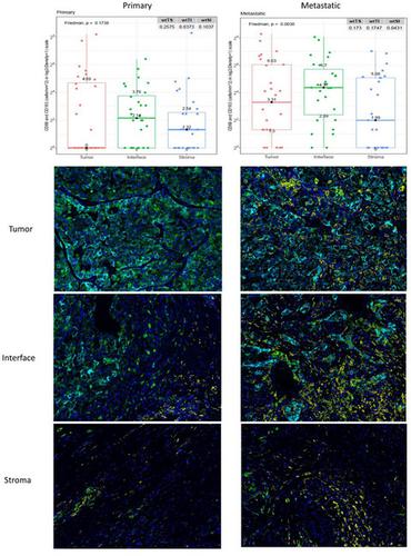
A clearer understanding of the tumor immune microenvironment (TIME) in metastatic clear cell renal cell carcinoma (ccRCC) may help to inform precision treatment strategies. We sought to identify clinically meaningful TIME signatures in ccRCC. We studied tumors from 39 patients with metastatic ccRCC using quantitative multiplexed immunofluorescence and relevant immune marker panels. Cell densities were analyzed in three regions of interest (ROIs): tumor core, tumor–stroma interface and stroma. Patients were stratified into low‐ and high‐marker density groups using median values as thresholds. Log‐rank and Cox regression analyses while controlling for clinical variables were used to compare survival outcomes to patterns of immune cell distributions. There were significant associations with increased macrophage (CD68+CD163+CD206+) density and poor outcomes across multiple ROIs in primary and metastatic tumors. In primary tumors, T‐bet+ T helper type 1 (Th1) cell density was highest at the tumor–stromal interface (P = 0·0021), and increased co‐expression of CD3 and T‐bet was associated with improved overall survival (P = 0·015) and survival after immunotherapy (P = 0·014). In metastatic tumor samples, decreased forkhead box protein 3 (FoxP3)+ T regulatory cell density correlated with improved survival after immunotherapy (P = 0·016). Increased macrophage markers and decreased Th1 T cell markers within the TIME correlated with poor overall survival and treatment outcomes. Immune markers such as FoxP3 showed consistent levels across the TIME, whereas others, such as T‐bet, demonstrated significant variance across the distinct ROIs. These findings suggest that TIME profiling outside the tumor core may identify clinically relevant associations for patients with metastatic ccRCC.
中文翻译:

转移性肾细胞癌肿瘤免疫微环境空间异质性及其与免疫治疗反应的相关性
更清楚地了解转移性透明细胞肾细胞癌 (ccRCC) 中的肿瘤免疫微环境 (TIME) 可能有助于制定精准治疗策略。我们试图确定 ccRCC 中具有临床意义的 TIME 特征。我们使用定量多重免疫荧光和相关免疫标记组研究了来自 39 名转移性 ccRCC 患者的肿瘤。在三个感兴趣区域(ROI)中分析细胞密度:肿瘤核心、肿瘤-基质界面和基质。使用中值作为阈值将患者分层为低和高标志物密度组。在控制临床变量的同时,使用对数秩和 Cox 回归分析来比较生存结果与免疫细胞分布模式。与巨噬细胞(CD68 +CD163 + CD206 + ) 原发性和转移性肿瘤中多个 ROI 的密度和不良结果。在原发性肿瘤中,T-bet + T 辅助 1 型 (Th1) 细胞密度在肿瘤-基质界面处最高(P = 0·0021),CD3 和 T-bet 共表达增加与总生存期改善相关( P = 0·015) 和免疫治疗后的生存率 ( P = 0·014)。在转移性肿瘤样本中,叉头盒蛋白 3 (FoxP3) + T 调节细胞密度降低与免疫治疗后存活率提高相关(P = 0·016)。TIME 内巨噬细胞标志物增加和 Th1 T 细胞标志物减少与较差的总体存活率和治疗结果相关。FoxP3 等免疫标记在整个 TIME 中显示出一致的水平,而其他免疫标记,例如 T-bet,在不同的 ROI 中表现出显着差异。这些发现表明,肿瘤核心以外的时间分析可以确定转移性 ccRCC 患者的临床相关关联。
更新日期:2020-12-21
中文翻译:
转移性肾细胞癌肿瘤免疫微环境空间异质性及其与免疫治疗反应的相关性
更清楚地了解转移性透明细胞肾细胞癌 (ccRCC) 中的肿瘤免疫微环境 (TIME) 可能有助于制定精准治疗策略。我们试图确定 ccRCC 中具有临床意义的 TIME 特征。我们使用定量多重免疫荧光和相关免疫标记组研究了来自 39 名转移性 ccRCC 患者的肿瘤。在三个感兴趣区域(ROI)中分析细胞密度:肿瘤核心、肿瘤-基质界面和基质。使用中值作为阈值将患者分层为低和高标志物密度组。在控制临床变量的同时,使用对数秩和 Cox 回归分析来比较生存结果与免疫细胞分布模式。与巨噬细胞(CD68 +CD163 + CD206 + ) 原发性和转移性肿瘤中多个 ROI 的密度和不良结果。在原发性肿瘤中,T-bet + T 辅助 1 型 (Th1) 细胞密度在肿瘤-基质界面处最高(P = 0·0021),CD3 和 T-bet 共表达增加与总生存期改善相关( P = 0·015) 和免疫治疗后的生存率 ( P = 0·014)。在转移性肿瘤样本中,叉头盒蛋白 3 (FoxP3) + T 调节细胞密度降低与免疫治疗后存活率提高相关(P = 0·016)。TIME 内巨噬细胞标志物增加和 Th1 T 细胞标志物减少与较差的总体存活率和治疗结果相关。FoxP3 等免疫标记在整个 TIME 中显示出一致的水平,而其他免疫标记,例如 T-bet,在不同的 ROI 中表现出显着差异。这些发现表明,肿瘤核心以外的时间分析可以确定转移性 ccRCC 患者的临床相关关联。


 京公网安备 11010802027423号