当前位置:
X-MOL 学术
›
J. Appl. Crystallogr.
›
论文详情
Our official English website, www.x-mol.net, welcomes your
feedback! (Note: you will need to create a separate account there.)
Optimizing laboratory X-ray diffraction contrast tomography for grain structure characterization of pure iron
Journal of Applied Crystallography ( IF 5.2 ) Pub Date : 2021-02-01 , DOI: 10.1107/s1600576720014673 Adam Lindkvist 1 , Haixing Fang 1 , Dorte Juul Jensen 1 , Yubin Zhang 1
Journal of Applied Crystallography ( IF 5.2 ) Pub Date : 2021-02-01 , DOI: 10.1107/s1600576720014673 Adam Lindkvist 1 , Haixing Fang 1 , Dorte Juul Jensen 1 , Yubin Zhang 1
Affiliation
|
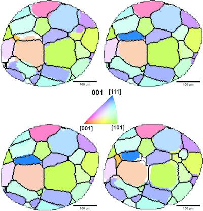
Laboratory diffraction contrast tomography (LabDCT) is a recently developed technique for 3D nondestructive grain mapping using a conical polychromatic beam from a laboratory-based X-ray source. The effects of experimental parameters, including accelerating voltage, exposure time and number of projections used for reconstruction, on the characterization of the 3D grain structure in an iron sample are quantified. The experiments were conducted using a commercial X-ray tomography system, ZEISS Xradia 520 Versa, equipped with a LabDCT module; and the data analysis was performed using the software package GrainMapper3D, which produces a 3D reconstruction from binarized 2D diffraction patterns. It is found that the exposure time directly affects the background noise level and thus the ability to distinguish weak spots of small grains from the background. With the assistance of forward simulations, it is found that spots from the first three strongest {hkl} families of a large grain can be seen with as few as 30–40 projections, which is sufficient for indexing the crystallographic orientation and resolving the grain shape with a reasonably high accuracy. It is also shown that the electron current is a more important factor than the accelerating voltage to be considered for optimizing the photon numbers with energies in the range of 20–60 keV. This energy range is the most important one for diffraction of common metals, e.g. iron and aluminium. Several suggestions for optimizing LabDCT experiments and 3D volume reconstruction are finally provided.
中文翻译:

优化实验室 X 射线衍射对比断层扫描以表征纯铁的晶粒结构
实验室衍射对比断层扫描 (LabDCT) 是最近开发的一项技术,使用来自实验室 X 射线源的锥形多色光束进行 3D 无损颗粒测绘。量化了实验参数(包括加速电压、曝光时间和用于重建的投影数量)对铁样品中 3D 晶粒结构表征的影响。实验使用配备 LabDCT 模块的商用 X 射线断层扫描系统 ZEISS Xradia 520 Versa 进行;数据分析是使用软件包 GrainMapper3D 进行的,该软件包可以根据二值化的 2D 衍射图样进行 3D 重建。研究发现,曝光时间直接影响背景噪声水平,从而影响区分小颗粒薄弱点与背景的能力。在正向模拟的帮助下,发现大晶粒的前三个最强的{hkl}族的斑点只需30-40个投影就可以看到,这足以索引晶体取向并解析晶粒形状具有相当高的准确度。研究还表明,在优化能量在 20-60 keV 范围内的光子数时,电子电流是比加速电压更重要的因素。该能量范围对于铁和铝等常见金属的衍射来说是最重要的。最后提出了优化 LabDCT 实验和 3D 体积重建的几点建议。
更新日期:2021-02-01
中文翻译:
优化实验室 X 射线衍射对比断层扫描以表征纯铁的晶粒结构
实验室衍射对比断层扫描 (LabDCT) 是最近开发的一项技术,使用来自实验室 X 射线源的锥形多色光束进行 3D 无损颗粒测绘。量化了实验参数(包括加速电压、曝光时间和用于重建的投影数量)对铁样品中 3D 晶粒结构表征的影响。实验使用配备 LabDCT 模块的商用 X 射线断层扫描系统 ZEISS Xradia 520 Versa 进行;数据分析是使用软件包 GrainMapper3D 进行的,该软件包可以根据二值化的 2D 衍射图样进行 3D 重建。研究发现,曝光时间直接影响背景噪声水平,从而影响区分小颗粒薄弱点与背景的能力。在正向模拟的帮助下,发现大晶粒的前三个最强的{hkl}族的斑点只需30-40个投影就可以看到,这足以索引晶体取向并解析晶粒形状具有相当高的准确度。研究还表明,在优化能量在 20-60 keV 范围内的光子数时,电子电流是比加速电压更重要的因素。该能量范围对于铁和铝等常见金属的衍射来说是最重要的。最后提出了优化 LabDCT 实验和 3D 体积重建的几点建议。


 京公网安备 11010802027423号