Cellular and Molecular Gastroenterology and Hepatology ( IF 7.1 ) Pub Date : 2020-12-16 , DOI: 10.1016/j.jcmgh.2020.12.007 Tingting Su 1 , Yilin Yang 2 , Sanchuan Lai 2 , Jain Jeong 2 , Yirang Jung 2 , Matthew McConnell 2 , Teruo Utsumi 2 , Yasuko Iwakiri 2
|
Background
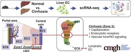
Dysfunction of liver sinusoidal endothelial cells (LSECs) is permissive for the progression of liver fibrosis and cirrhosis and responsible for its clinical complications. Here, we have mapped the spatial distribution of heterogeneous liver ECs in normal vs cirrhotic mouse livers and identified zone-specific transcriptomic changes of LSECs associated with liver cirrhosis using scRNA-seq technology.
Approach & Results
Cirrhosis was generated in endothelial specific green fluorescent protein (GFP) reporter mice through carbon tetrachloride inhalation for 12 weeks. GFP-positive liver EC populations were isolated from control and cirrhotic mice by FACS. We identified 6 clusters of liver EC populations including 3 clusters of LSECs, 2 clusters of vascular ECs and 1 cluster of lymphatic ECs. Based on previously reported LSEC-landmarks, we mapped the 3 clusters of LSECs in zones 1, 2, and 3, and determined phenotypic changes in each zone between control and cirrhotic mice. We found genes representing capillarization of LSECs (eg, CD34) as well as extracellular matrix genes were most upregulated in LSECs of zone 3 in cirrhotic mice, which may contribute to the development of basement membranes. LSECs in cirrhotic mice also demonstrated decreased expression of endocytic receptors, most remarkably in zone 3. Transcription factors (Klf2 [Kruppel-like factor-2], Klf4 [Kruppel-like factor-4], and AP-1) that induce nitric oxide production in response to shear stress were downregulated in LSECs of all zones in cirrhotic mice, implying increased intrahepatic vascular resistance.
Conclusion
This study deepens our knowledge of the pathogenesis of liver cirrhosis at a spatial, cell-specific level, which is indispensable for the development of novel therapeutic strategies to target the most dysfunctional liver ECs.
中文翻译:
单细胞转录组学揭示肝硬化中肝窦内皮细胞的区域特异性改变
背景
肝窦内皮细胞(LSEC)功能障碍导致肝纤维化和肝硬化的进展,并导致其临床并发症。在这里,我们绘制了正常小鼠肝脏与肝硬化小鼠肝脏中异质肝脏 EC 的空间分布,并使用 scRNA-seq 技术识别了与肝硬化相关的 LSEC 的区域特异性转录组变化。
方法与结果
通过吸入四氯化碳12周,使内皮特异性绿色荧光蛋白(GFP)报告小鼠产生肝硬化。通过 FACS 从对照小鼠和肝硬化小鼠中分离出 GFP 阳性肝脏 EC 群体。我们确定了 6 个肝脏 EC 群体簇,包括 3 个 LSEC 簇、2 个血管 EC 簇和 1 个淋巴 EC 簇。根据先前报道的 LSEC 标志,我们绘制了 1、2 和 3 区中的 3 个 LSEC 簇,并确定了对照小鼠和肝硬化小鼠之间每个区域的表型变化。我们发现代表 LSEC 毛细血管化的基因(例如 CD34)以及细胞外基质基因在肝硬化小鼠第 3 区的 LSEC 中上调最多,这可能有助于基底膜的发育。肝硬化小鼠的 LSEC 也表现出内吞受体表达下降,最明显的是在 3 区。诱导一氧化氮的转录因子(Klf2 [Kruppel 样因子 2]、Klf4 [Kruppel 样因子 4] 和 AP-1)肝硬化小鼠所有区域的 LSEC 中响应剪切应力的生成量均下调,这意味着肝内血管阻力增加。
结论
这项研究加深了我们在空间、细胞特异性水平上对肝硬化发病机制的认识,这对于开发针对功能最差的肝脏内皮细胞的新型治疗策略是必不可少的。


 京公网安备 11010802027423号