Experimental Cell Research ( IF 3.3 ) Pub Date : 2020-12-16 , DOI: 10.1016/j.yexcr.2020.112434 Rafaela Rossetti , Felipe Augusto Rós , Lucas Eduardo Botelho de Souza , Juliana de Matos Maçonetto , Péricles Natan Mendes da Costa , Fernanda Ursoli Ferreira , Josiane Serrano Borges , Julianne Vargas de Carvalho , Nayara Patrícia Morotti , Simone Kashima , Dimas Tadeu Covas
|
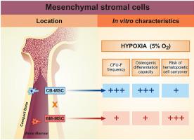
It has been suggested that the bone marrow microenvironment harbors two distinct populations of mesenchymal stromal cells (MSC), one with a perivascular location and other present in the endosteum. A better understanding of the biology of these MSC subsets has been pursued in order to refine its clinical application. However, most comparative characterizations of mouse MSC have been performed in normoxia. This can result in misleading interpretations since mouse MSC subsets with low/defective p53 activity are known to be selected during culture in normoxia. Here, we report a comprehensive in vitro characterization of mouse MSC isolated from bone marrow (BM-MSC) and compact bone (CB-MSC) expanded and assayed under hypoxia for their morphology, clonogenic efficiency and differentiation capacity. We found that, under hypoxia, compact bone is richer in absolute numbers of MSC and isolation of MSC from compact bone is associated with a reduced risk of hematopoietic cell carryover. In addition, CB-MSC have higher in vitro osteogenic capacity than BM-MSC, while adipogenic differentiation potential is similar. These findings reinforce the hypothesis of the existence of MSC in bone marrow and compact bone representing functionally distinct cell populations and highlight the compact bone as an efficient source of murine MSC under physiological oxygen concentrations.
中文翻译:
缺氧培养的小鼠骨髓间质基质细胞和致密骨表现出不同的表型特征
已经提出,骨髓微环境具有两个不同的间充质基质细胞(MSC)群体,一个具有血管周围位置,另一个存在于内膜中。为了完善其临床应用,已经寻求对这些MSC亚组的生物学的更好理解。但是,大多数小鼠MSC的比较特征是在常氧状态下进行的。这可能导致误导性的解释,因为已知在常氧培养过程中选择了p53活性低/有缺陷的小鼠MSC亚群。在这里,我们报告了全面的体外小鼠骨髓间充质干细胞(BM-MSC)和致密骨(CB-MSC)的扩展特性,并在缺氧条件下对其形态,克隆形成效率和分化能力进行了测定。我们发现,在缺氧的情况下,致密骨中的MSC绝对数量更丰富,从致密骨中分离MSC与降低造血细胞残留的风险有关。此外,CB-MSC的体外成骨能力高于BM-MSC,而成脂分化潜能相似。这些发现加强了在代表功能上不同的细胞群的骨髓和紧密骨中存在MSC的假设,并强调了紧密骨在生理氧气浓度下作为鼠MSC的有效来源。


 京公网安备 11010802027423号