当前位置:
X-MOL 学术
›
Microsc. Res. Tech.
›
论文详情
Our official English website, www.x-mol.net, welcomes your
feedback! (Note: you will need to create a separate account there.)
The fine structure of the turbot eye (Scophtalmus maximus): A macro-anatomical, light and scanning electron microscopical study
Microscopy Research and Technique ( IF 2.0 ) Pub Date : 2020-12-14 , DOI: 10.1002/jemt.23674 Burcu Onuk 1 , Ozgür Y Pehlivan 2 , Banu Yardimci 3
Microscopy Research and Technique ( IF 2.0 ) Pub Date : 2020-12-14 , DOI: 10.1002/jemt.23674 Burcu Onuk 1 , Ozgür Y Pehlivan 2 , Banu Yardimci 3
Affiliation
|
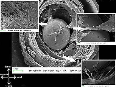
In fish species, the morphological structure of the eye varies depending on environmental conditions. Morphometric data about the sensory organs of fish is lacking. Therefore, this study aims to describe the morphological structure of the turbot eye using gross, light and scanning electron microscope examinations. The turbot eyeball was found to comprise three layers: the tunica fibrosa bulbi (cornea, sclera), the tunica vasculosa bulbi (choroidea, iris) and the tunica nervea bulbi (retina). The thickness of the centre of the cornea measured approximately 153.14 μm, and the peripheral thickness measured 410.81 μm. The sclera consisted of a two-part cartilage structure that was connected with elastic fibres. The choroideal rete was found in the tunica vasculosa bulbi, and its thickness measured 1.6 ± 0.1 mm. Moreover, no pigment was found in the choroidea. The lens was determined to be a very hard and transparent structure extending towards the cornea. In addition, we detected five ligaments in the equatorial plane of the eye, in which the tendon of the retractor lentis muscle attaches to the lens. Also, there were six extraocular muscles in the turbot. This study is the first to present detailed descriptions of morphological structures and morphometric data for all the layers of the turbot eye. Since the anatomical structure of the eye in fish is variable, it is thought that the data on the turbot eye will contribute to the anatomy literature.
中文翻译:

大菱鲆(Scophtalmus maximus)的精细结构:宏观解剖、光和扫描电子显微镜研究
在鱼类中,眼睛的形态结构因环境条件而异。缺乏有关鱼类感觉器官的形态学数据。因此,本研究旨在通过大体、光学和扫描电子显微镜检查来描述大菱鲆眼的形态结构。大菱鲆眼球由三层组成:球膜纤维(角膜、巩膜)、球膜血管(脉络膜、虹膜)和球神经膜(视网膜)。角膜中心厚度约为 153.14 μm,周边厚度为 410.81 μm。巩膜由两部分软骨结构组成,软骨结构与弹性纤维相连。脉络膜网位于血管球囊中,其厚度为 1.6 ± 0.1 mm。而且,在脉络膜中未发现色素。晶状体被确定是一个非常坚硬和透明的结构,向角膜延伸。此外,我们在眼睛的赤道平面上检测到 5 条韧带,其中晶状体牵开肌的肌腱附着在晶状体上。此外,大菱鲆有六块眼外肌。这项研究首次详细描述了大菱鲆眼睛所有层的形态结构和形态测量数据。由于鱼眼睛的解剖结构是可变的,因此认为大菱鲆眼睛的数据将对解剖学文献有所贡献。其中牵开晶状体肌的肌腱附着在晶状体上。此外,大菱鲆有六块眼外肌。这项研究首次详细描述了大菱鲆眼睛所有层的形态结构和形态测量数据。由于鱼眼睛的解剖结构是可变的,因此认为大菱鲆眼睛的数据将对解剖学文献有所贡献。其中牵开晶状体肌的肌腱附着在晶状体上。此外,大菱鲆有六块眼外肌。这项研究首次详细描述了大菱鲆眼睛所有层的形态结构和形态测量数据。由于鱼眼睛的解剖结构是可变的,因此认为大菱鲆眼睛的数据将对解剖学文献有所贡献。
更新日期:2020-12-14
中文翻译:
大菱鲆(Scophtalmus maximus)的精细结构:宏观解剖、光和扫描电子显微镜研究
在鱼类中,眼睛的形态结构因环境条件而异。缺乏有关鱼类感觉器官的形态学数据。因此,本研究旨在通过大体、光学和扫描电子显微镜检查来描述大菱鲆眼的形态结构。大菱鲆眼球由三层组成:球膜纤维(角膜、巩膜)、球膜血管(脉络膜、虹膜)和球神经膜(视网膜)。角膜中心厚度约为 153.14 μm,周边厚度为 410.81 μm。巩膜由两部分软骨结构组成,软骨结构与弹性纤维相连。脉络膜网位于血管球囊中,其厚度为 1.6 ± 0.1 mm。而且,在脉络膜中未发现色素。晶状体被确定是一个非常坚硬和透明的结构,向角膜延伸。此外,我们在眼睛的赤道平面上检测到 5 条韧带,其中晶状体牵开肌的肌腱附着在晶状体上。此外,大菱鲆有六块眼外肌。这项研究首次详细描述了大菱鲆眼睛所有层的形态结构和形态测量数据。由于鱼眼睛的解剖结构是可变的,因此认为大菱鲆眼睛的数据将对解剖学文献有所贡献。其中牵开晶状体肌的肌腱附着在晶状体上。此外,大菱鲆有六块眼外肌。这项研究首次详细描述了大菱鲆眼睛所有层的形态结构和形态测量数据。由于鱼眼睛的解剖结构是可变的,因此认为大菱鲆眼睛的数据将对解剖学文献有所贡献。其中牵开晶状体肌的肌腱附着在晶状体上。此外,大菱鲆有六块眼外肌。这项研究首次详细描述了大菱鲆眼睛所有层的形态结构和形态测量数据。由于鱼眼睛的解剖结构是可变的,因此认为大菱鲆眼睛的数据将对解剖学文献有所贡献。


 京公网安备 11010802027423号