Immunity ( IF 32.4 ) Pub Date : 2020-12-14 , DOI: 10.1016/j.immuni.2020.12.002 Min-Seok Rha , Hye Won Jeong , Jae-Hoon Ko , Seong Jin Choi , In-Ho Seo , Jeong Seok Lee , Moa Sa , A Reum Kim , Eun-Jeong Joo , Jin Young Ahn , Jung Ho Kim , Kyoung-Ho Song , Eu Suk Kim , Dong Hyun Oh , Mi Young Ahn , Hee Kyoung Choi , Ji Hoon Jeon , Jae-Phil Choi , Hong Bin Kim , Young Keun Kim , Su-Hyung Park , Won Suk Choi , Jun Yong Choi , Kyong Ran Peck , Eui-Cheol Shin
|
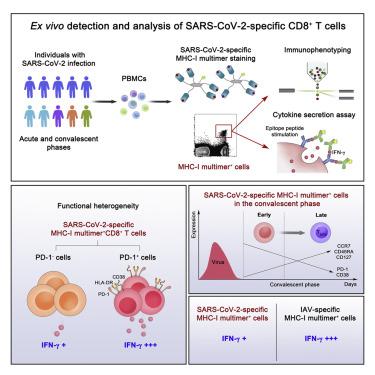
Memory T cell responses have been demonstrated in COVID-19 convalescents, but ex vivo phenotypes of SARS-CoV-2-specific T cells have been unclear. We detected SARS-CoV-2-specific CD8+ T cells by MHC class I multimer staining and examined their phenotypes and functions in acute and convalescent COVID-19. Multimer+ cells exhibited early differentiated effector-memory phenotypes in the early convalescent phase. The frequency of stem-like memory cells was increased among multimer+ cells in the late convalescent phase. Cytokine secretion assays combined with MHC class I multimer staining revealed that the proportion of interferon-γ (IFN-γ)-producing cells was significantly lower among SARS-CoV-2-specific CD8+ T cells than those specific to influenza A virus. Importantly, the proportion of IFN-γ-producing cells was higher in PD-1+ cells than PD-1− cells among multimer+ cells, indicating that PD-1-expressing, SARS-CoV-2-specific CD8+ T cells are not exhausted, but functional. Our current findings provide information for understanding of SARS-CoV-2-specific CD8+ T cells elicited by infection or vaccination.
中文翻译:
PD-1表达SARS-CoV-2特异性CD8 + T细胞并未耗尽,但在COVID-19患者中起作用
已在COVID-19恢复期中证明了记忆T细胞反应,但SARS-CoV-2特异性T细胞的离体表型尚不清楚。我们通过MHC I类多聚体染色检测到SARS-CoV-2特异性CD8 + T细胞,并检查了它们在急性和恢复期COVID-19中的表型和功能。在恢复期早期,多聚体+细胞表现出早期分化的效应记忆表型。在恢复期后期,多聚体+细胞中干细胞样记忆细胞的频率增加。细胞因子分泌测定结合MHC I类多聚体染色显示,在SARS-CoV-2特异性CD8 +中,产生干扰素-γ(IFN-γ)的细胞比例明显降低T细胞比那些甲型流感病毒特异的。重要的是,IFN-γ产生细胞的比例是在PD-1更高+细胞中比PD-1 -细胞多聚体间+细胞,这表明PD-1表达,SARS-CoV的-2特异性CD8 + T细胞是不是精疲力尽,而是功能正常。我们当前的发现为理解感染或接种疫苗引起的SARS-CoV-2特异性CD8 + T细胞提供了信息。


 京公网安备 11010802027423号