Neuroscience Research ( IF 2.4 ) Pub Date : 2020-12-11 , DOI: 10.1016/j.neures.2020.11.004 Maria Izco 1 , Javier Blesa 2 , Guglielmo Verona 3 , J Mark Cooper 4 , Lydia Alvarez-Erviti 1
|
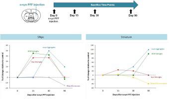
Neuroinflammation is increasingly recognized as an important feature in the pathogenesis of Parkinson’s disease (PD). However, it remains unclear whether neuroinflammation contributes to nigral degeneration in PD or is merely a secondary marker of neurodegeneration. We aimed to investigate the temporal relationship between synucleopathy, neuroinflammation and nigrostriatal degeneration in a mouse model of PD. Mice received unilateral intrastriatal injection of alpha-synuclein pre-formed fibrils, alpha-synuclein monomer or vehicle and were sacrificed at 15, 30 and 90 days post-injection. Intrastriatal inoculation of alpha-synuclein fibrils led to significant alpha-synuclein aggregation in the substantia nigra peaking at 30 days after injection while the significant increase in Iba-1 cells, GFAP cells and IL-1β expression peaked earlier at 15 days. At 90 days, the striatal dopaminergic denervation was associated with astroglial activation. Alpha-synuclein monomer did not result in long-term glia activation or increase in inflammatory markers. The spread of alpha-synuclein aggregates into the cortex was not associated with any changes to neuroinflammatory markers. Our results demonstrate that in the substantia nigra glial activation is an early event that precedes alpha-synuclein inclusion formation, suggesting neuroinflammation could play an important early role in the pathogenesis of PD.
中文翻译:
在帕金森病小鼠模型中,胶质细胞激活先于 α-突触核蛋白病理学
神经炎症越来越被认为是帕金森病 (PD) 发病机制中的一个重要特征。然而,尚不清楚神经炎症是否会导致 PD 中的黑质变性,或者仅仅是神经变性的次要标志物。我们旨在研究 PD 小鼠模型中突触核病、神经炎症和黑质纹状体变性之间的时间关系。小鼠接受α-突触核蛋白预制原纤维、α-突触核蛋白单体或载体的单侧纹状体内注射,并在注射后15、30和90天处死。α-突触核蛋白原纤维的纹状体接种导致黑质中的显着α-突触核蛋白聚集在注射后30天达到峰值,而Iba-1细胞、GFAP细胞和IL-1β表达的显着增加在15天达到峰值。在 90 天时,纹状体多巴胺能去神经支配与星形胶质细胞激活有关。α-突触核蛋白单体不会导致长期胶质细胞激活或炎症标志物增加。α-突触核蛋白聚集体扩散到皮层与神经炎症标志物的任何变化无关。我们的研究结果表明,在黑质中,神经胶质激活是α-突触核蛋白包涵体形成之前的早期事件,这表明神经炎症可能在 PD 的发病机制中发挥重要的早期作用。α-突触核蛋白聚集体扩散到皮层与神经炎症标志物的任何变化无关。我们的研究结果表明,在黑质中,神经胶质激活是α-突触核蛋白包涵体形成之前的早期事件,这表明神经炎症可能在 PD 的发病机制中发挥重要的早期作用。α-突触核蛋白聚集体扩散到皮层与神经炎症标志物的任何变化无关。我们的研究结果表明,在黑质中,神经胶质激活是α-突触核蛋白包涵体形成之前的早期事件,这表明神经炎症可能在 PD 的发病机制中发挥重要的早期作用。


 京公网安备 11010802027423号