当前位置:
X-MOL 学术
›
Biotechnol. Bioeng.
›
论文详情
Our official English website, www.x-mol.net, welcomes your
feedback! (Note: you will need to create a separate account there.)
In situ tracking of microbeads for the detection of biofilm formation
Biotechnology and Bioengineering ( IF 3.5 ) Pub Date : 2020-12-10 , DOI: 10.1002/bit.27648 Héloïse Boudarel 1 , Jean-Denis Mathias 2 , Benoît Blaysat 1 , Michel Grédiac 1
Biotechnology and Bioengineering ( IF 3.5 ) Pub Date : 2020-12-10 , DOI: 10.1002/bit.27648 Héloïse Boudarel 1 , Jean-Denis Mathias 2 , Benoît Blaysat 1 , Michel Grédiac 1
Affiliation
|
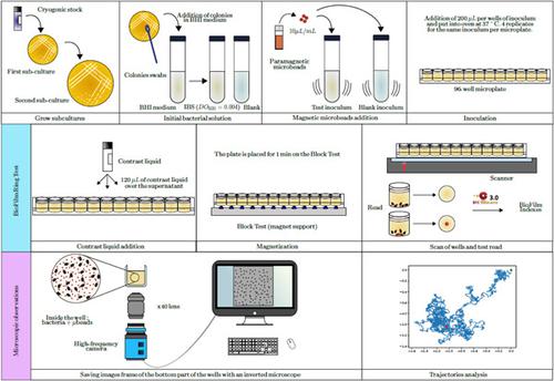
In this study, we utilize the free motion of beads incorporated in bacterial suspension to investigate the behavior of the medium surrounding the beads during biofilm formation. The use of imaging techniques such as digital image correlation enables tracking of the movement of beads, which serve as markers in the processed images. This method is applied to detect and characterize biofilm formation. The main originality of this study lies in characterizing the evolution of the typology of bead movements during biofilm formation. The aim is to identify bead behaviors that represent the start of biofilm formation. By observing inert bead movements introduced into the bacterial environment, changes in trajectory typologies are detected and appear to be related to sessile bacterial activity, bacterial hindrance, and adhesion or formation of extracellular material. We use our approach to discriminate between the presence or absence of antibiotics mixed with bacteria and to assess their effectiveness. The results highlight the potential of our approach as nondestructive tracking of biofilm dynamics over time based on optical microscope images.
中文翻译:

用于检测生物膜形成的微珠原位跟踪
在这项研究中,我们利用细菌悬浮液中珠子的自由运动来研究生物膜形成过程中珠子周围介质的行为。使用数字图像相关等成像技术可以跟踪珠子的运动,这些珠子在处理后的图像中充当标记。该方法用于检测和表征生物膜的形成。本研究的主要原创性在于描述生物膜形成过程中珠粒运动类型的演变。目的是确定代表生物膜形成开始的珠行为。通过观察引入细菌环境的惰性珠粒运动,可以检测到轨迹类型的变化,并且似乎与固着细菌活动、细菌阻碍、和细胞外物质的粘附或形成。我们使用我们的方法来区分是否存在与细菌混合的抗生素并评估其有效性。结果突出了我们的方法作为基于光学显微镜图像随时间推移对生物膜动力学进行非破坏性跟踪的潜力。
更新日期:2021-02-18
中文翻译:
用于检测生物膜形成的微珠原位跟踪
在这项研究中,我们利用细菌悬浮液中珠子的自由运动来研究生物膜形成过程中珠子周围介质的行为。使用数字图像相关等成像技术可以跟踪珠子的运动,这些珠子在处理后的图像中充当标记。该方法用于检测和表征生物膜的形成。本研究的主要原创性在于描述生物膜形成过程中珠粒运动类型的演变。目的是确定代表生物膜形成开始的珠行为。通过观察引入细菌环境的惰性珠粒运动,可以检测到轨迹类型的变化,并且似乎与固着细菌活动、细菌阻碍、和细胞外物质的粘附或形成。我们使用我们的方法来区分是否存在与细菌混合的抗生素并评估其有效性。结果突出了我们的方法作为基于光学显微镜图像随时间推移对生物膜动力学进行非破坏性跟踪的潜力。


 京公网安备 11010802027423号