当前位置:
X-MOL 学术
›
J. Neurosci. Res.
›
论文详情
Our official English website, www.x-mol.net, welcomes your
feedback! (Note: you will need to create a separate account there.)
Conventional immunomarkers stain a fraction of astrocytes in vitro: A comparison of rat cortical and spinal cord astrocytes in naïve and stimulated cultures
Journal of Neuroscience Research ( IF 2.9 ) Pub Date : 2020-12-08 , DOI: 10.1002/jnr.24759 Bailey Balouch 1, 2, 3 , Jessica L Funnell 1, 2 , Alexis M Ziemba 1, 2, 4 , Devan L Puhl 1, 2 , Kathy Lin 1, 2 , Manoj K Gottipati 1, 2, 5 , Ryan J Gilbert 1, 2
Journal of Neuroscience Research ( IF 2.9 ) Pub Date : 2020-12-08 , DOI: 10.1002/jnr.24759 Bailey Balouch 1, 2, 3 , Jessica L Funnell 1, 2 , Alexis M Ziemba 1, 2, 4 , Devan L Puhl 1, 2 , Kathy Lin 1, 2 , Manoj K Gottipati 1, 2, 5 , Ryan J Gilbert 1, 2
Affiliation
|
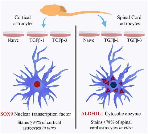
Astrocytes are responsible for a wide variety of essential functions throughout the central nervous system. The protein markers glial fibrillary acidic protein (GFAP), glutamate aspartate transporter (GLAST), glutamate transporter‐1 (GLT‐1), glutamine synthetase (GS), 10‐formyltetrahydrofolate dehydrogenase (ALDH1L1), and the transcription factor SOX9 are routinely used to label astrocytes in primary rodent cultures. However, GLAST, GLT‐1, GS, and SOX9 are also produced by microglia and oligodendrocytes and GFAP, GLAST, GLT‐1, and GS production levels are affected by astrocyte phenotypic changes associated with reactive astrogliosis. No group has performed a comprehensive immunocytochemical evaluation to quantify the percentage of cells labeled by these markers in vitro, nor compared changes in staining between cortex‐ and spinal cord‐derived cells in naïve and stimulated cultures. Here, we quantified the percentage of cells positively stained for these six markers in astrocyte, microglia, and oligodendrocyte cultures isolated from neonatal rat cortices and spinal cords. Additionally, we incubated the astrocytes with transforming growth factor (TGF)‐β1 or TGF‐β3 to determine if the labeling of these markers is altered by these stimuli. We found that only SOX9 in cortical cultures and ALDH1L1 in spinal cord cultures labeled more than 75% of the cells in naïve and stimulated astrocyte cultures and stained less than 5% of the cells in microglia and oligodendrocyte cultures. Furthermore, significantly more cortical than spinal cord astrocytes stained for GFAP, GLAST, and ALDH1L1 in naïve cultures, whereas significantly more spinal cord than cortical astrocytes stained for GLAST and GS in TGF‐β1‐treated cultures. These findings are important as variability in marker staining may lead to misinterpretation of the astrocyte response in cocultures, migration assays, or engineered disease models.
中文翻译:

常规免疫标志物在体外对一部分星形胶质细胞染色:幼稚和刺激培养物中大鼠皮质和脊髓星形胶质细胞的比较
星形胶质细胞负责整个中枢神经系统的多种基本功能。蛋白质标记物胶质纤维酸性蛋白 (GFAP)、谷氨酸天冬氨酸转运蛋白 (GLAST)、谷氨酸转运蛋白-1 (GLT-1)、谷氨酰胺合成酶 (GS)、10-甲酰基四氢叶酸脱氢酶 (ALDH1L1) 和转录因子 SOX9 是常规使用的在初级啮齿动物培养物中标记星形胶质细胞。然而,GLAST、GLT-1、GS 和 SOX9 也由小胶质细胞和少突胶质细胞产生,GFAP、GLAST、GLT-1 和 GS 的产生水平受与反应性星形胶质细胞增生相关的星形胶质细胞表型变化的影响。没有一个小组进行过全面的免疫细胞化学评估来量化体外这些标记物标记的细胞百分比,也没有比较幼稚和刺激培养物中皮层和脊髓衍生细胞之间染色的变化。在这里,我们量化了从新生大鼠皮质和脊髓中分离出的星形胶质细胞、小胶质细胞和少突胶质细胞培养物中这六种标记物阳性染色的细胞百分比。此外,我们将星形胶质细胞与转化生长因子 (TGF)-β1 或 TGF-β3 一起孵育,以确定这些标记物的标记是否因这些刺激而改变。我们发现,只有皮质培养物中的 SOX9 和脊髓培养物中的 ALDH1L1 标记了幼稚和刺激星形胶质细胞培养物中超过 75% 的细胞,并染色了小胶质细胞和少突胶质细胞培养物中不到 5% 的细胞。此外,在幼稚培养物中的 GFAP、GLAST 和 ALDH1L1 染色的皮质星形胶质细胞明显多于脊髓,而在 TGF-β1 处理的培养物中,脊髓的 GLAST 和 GS 染色明显多于皮质星形胶质细胞。这些发现很重要,因为标记染色的可变性可能导致对共培养、迁移测定或工程疾病模型中星形胶质细胞反应的误解。
更新日期:2021-01-29
中文翻译:
常规免疫标志物在体外对一部分星形胶质细胞染色:幼稚和刺激培养物中大鼠皮质和脊髓星形胶质细胞的比较
星形胶质细胞负责整个中枢神经系统的多种基本功能。蛋白质标记物胶质纤维酸性蛋白 (GFAP)、谷氨酸天冬氨酸转运蛋白 (GLAST)、谷氨酸转运蛋白-1 (GLT-1)、谷氨酰胺合成酶 (GS)、10-甲酰基四氢叶酸脱氢酶 (ALDH1L1) 和转录因子 SOX9 是常规使用的在初级啮齿动物培养物中标记星形胶质细胞。然而,GLAST、GLT-1、GS 和 SOX9 也由小胶质细胞和少突胶质细胞产生,GFAP、GLAST、GLT-1 和 GS 的产生水平受与反应性星形胶质细胞增生相关的星形胶质细胞表型变化的影响。没有一个小组进行过全面的免疫细胞化学评估来量化体外这些标记物标记的细胞百分比,也没有比较幼稚和刺激培养物中皮层和脊髓衍生细胞之间染色的变化。在这里,我们量化了从新生大鼠皮质和脊髓中分离出的星形胶质细胞、小胶质细胞和少突胶质细胞培养物中这六种标记物阳性染色的细胞百分比。此外,我们将星形胶质细胞与转化生长因子 (TGF)-β1 或 TGF-β3 一起孵育,以确定这些标记物的标记是否因这些刺激而改变。我们发现,只有皮质培养物中的 SOX9 和脊髓培养物中的 ALDH1L1 标记了幼稚和刺激星形胶质细胞培养物中超过 75% 的细胞,并染色了小胶质细胞和少突胶质细胞培养物中不到 5% 的细胞。此外,在幼稚培养物中的 GFAP、GLAST 和 ALDH1L1 染色的皮质星形胶质细胞明显多于脊髓,而在 TGF-β1 处理的培养物中,脊髓的 GLAST 和 GS 染色明显多于皮质星形胶质细胞。这些发现很重要,因为标记染色的可变性可能导致对共培养、迁移测定或工程疾病模型中星形胶质细胞反应的误解。


 京公网安备 11010802027423号