当前位置:
X-MOL 学术
›
Mol. Oral Microbiol.
›
论文详情
Our official English website, www.x-mol.net, welcomes your
feedback! (Note: you will need to create a separate account there.)
Aggregatibacter actinomycetemcomitans induces a proatherosclerotic response in human endothelial cells in a three‐dimensional collagen scaffold model
Molecular Oral Microbiology ( IF 2.8 ) Pub Date : 2020-12-07 , DOI: 10.1111/omi.12326 Maria A. Torres 1, 2 , Diego F. Gualtero 1, 2 , Gloria I. Lafaurie 2 , Marta R. Fontanilla 1
Molecular Oral Microbiology ( IF 2.8 ) Pub Date : 2020-12-07 , DOI: 10.1111/omi.12326 Maria A. Torres 1, 2 , Diego F. Gualtero 1, 2 , Gloria I. Lafaurie 2 , Marta R. Fontanilla 1
Affiliation
|
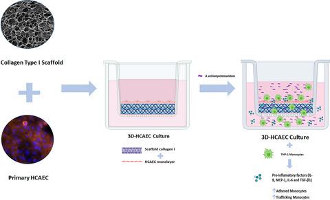
The role of periodontopathogens in inflammatory endothelial dysfunction is not known. This study characterizes a three‐dimensional model with human coronary artery endothelial cells on three‐dimensional (HCAEC‐3D) type I collagen scaffolds to evaluate whether infection with Aggregatibacter actinomycetemcomitans induces a proinflammatory response associated with atherosclerosis. The HCAEC‐3D culture was physicochemically characterized with regard to biocompatibility and barrier function. Then, the culture was infected with A. actinomycetemcomitans strain ATCC 29522 at multiplicities of infection (MOIs) of 1:1, 1:10, and 1:100. Cultures without infection and stimulated with A. actinomycetemcomitans lipopolysaccharide were used as controls. The secretion of soluble factors (IL‐6, IL‐1β, MCP‐1, RANTES, MIP‐1, IL‐8, IL‐1α, and TNF‐α) was evaluated via flow cytometry; TGF‐β1 was evaluated via enzyme‐linked immunosorbent assay (ELISA). The adhesion and migration of fluorescent human THP‐1 monocytes was evaluated. IL‐8, MCP‐1, and IL‐6 secretion increased in a dose‐dependent manner with A. actinomycetemcomitans infection and was significantly greater than that under control treatment. The concentration of TGF‐β1 was significantly higher at MOI 1:100 than in controls. Treatment of the 3D cultures with A. actinomycetemcomitans at different MOIs induced significant differences in the adhesion of monocytes to the endothelium compared to the control without infection. Lastly, conditioned media from 3D cultures treated with A. actinomycetemcomitans induced monocyte migration. The effects of IL‐8, MCP‐1, IL‐6, and TGF‐β1 on the endothelium indicate the ability of A. actinomycetemcomitans to induce an inflammatory response through a mechanism of monocyte adhesion and migration and endothelial dysfunction.
中文翻译:

在三维胶原蛋白支架模型中,放线杆菌聚合酶诱导人内皮细胞的粥样硬化反应
牙周病原体在炎性内皮功能障碍中的作用尚不清楚。这项研究的特征是在三维(HCAEC-3D)I型胶原蛋白支架上构建了具有人类冠状动脉内皮细胞的三维模型,以评估感染聚合放线菌的大鼠是否会诱发与动脉粥样硬化相关的促炎反应。HCAEC-3D培养物具有生物相容性和屏障功能的物理化学特征。然后,以1:1、1:10和1:100的感染复数(MOI)用放线放线杆菌ATCC 29522菌株感染培养物。无感染且用放线放线杆菌刺激的培养脂多糖用作对照。通过流式细胞仪评估可溶性因子(IL-6,IL-1β,MCP-1,RANTES,MIP-1,IL-8,IL-1α和TNF-α)的分泌; 通过酶联免疫吸附测定(ELISA)对TGF-β1进行了评估。评估了荧光人THP-1单核细胞的黏附和迁移。IL-8,MCP-1和IL-6的分泌随放线放线杆菌感染的增加而呈剂量依赖性,并且显着高于对照治疗。MOI 1:100时,TGF-β1的浓度显着高于对照组。用放线放线杆菌对3D培养物的处理与没有感染的对照相比,在不同MOIs下,在不同MOIs下诱导的单核细胞对内皮粘附的显着差异。最后,来自用放线放线杆菌处理的3D培养物的条件培养基诱导单核细胞迁移。IL-8,MCP-1,IL-6和TGF-β1对内皮细胞的影响表明放线放线杆菌通过单核细胞粘附,迁移和内皮功能障碍机制诱导炎症反应的能力。
更新日期:2020-12-07
中文翻译:
在三维胶原蛋白支架模型中,放线杆菌聚合酶诱导人内皮细胞的粥样硬化反应
牙周病原体在炎性内皮功能障碍中的作用尚不清楚。这项研究的特征是在三维(HCAEC-3D)I型胶原蛋白支架上构建了具有人类冠状动脉内皮细胞的三维模型,以评估感染聚合放线菌的大鼠是否会诱发与动脉粥样硬化相关的促炎反应。HCAEC-3D培养物具有生物相容性和屏障功能的物理化学特征。然后,以1:1、1:10和1:100的感染复数(MOI)用放线放线杆菌ATCC 29522菌株感染培养物。无感染且用放线放线杆菌刺激的培养脂多糖用作对照。通过流式细胞仪评估可溶性因子(IL-6,IL-1β,MCP-1,RANTES,MIP-1,IL-8,IL-1α和TNF-α)的分泌; 通过酶联免疫吸附测定(ELISA)对TGF-β1进行了评估。评估了荧光人THP-1单核细胞的黏附和迁移。IL-8,MCP-1和IL-6的分泌随放线放线杆菌感染的增加而呈剂量依赖性,并且显着高于对照治疗。MOI 1:100时,TGF-β1的浓度显着高于对照组。用放线放线杆菌对3D培养物的处理与没有感染的对照相比,在不同MOIs下,在不同MOIs下诱导的单核细胞对内皮粘附的显着差异。最后,来自用放线放线杆菌处理的3D培养物的条件培养基诱导单核细胞迁移。IL-8,MCP-1,IL-6和TGF-β1对内皮细胞的影响表明放线放线杆菌通过单核细胞粘附,迁移和内皮功能障碍机制诱导炎症反应的能力。


 京公网安备 11010802027423号