当前位置:
X-MOL 学术
›
NMR Biomed.
›
论文详情
Our official English website, www.x-mol.net, welcomes your feedback! (Note: you will need to create a separate account there.)
Resolving the omega‐3 methyl resonance with long echo time magnetic resonance spectroscopy in mouse adipose tissue at 9.4 T
NMR in Biomedicine ( IF 2.9 ) Pub Date : 2020-12-02 , DOI: 10.1002/nbm.4455 Clara J Fallone 1 , Anthony G Tessier 1, 2 , Catherine J Field 3 , Atiyah Yahya 1, 2
NMR in Biomedicine ( IF 2.9 ) Pub Date : 2020-12-02 , DOI: 10.1002/nbm.4455 Clara J Fallone 1 , Anthony G Tessier 1, 2 , Catherine J Field 3 , Atiyah Yahya 1, 2
Affiliation
|
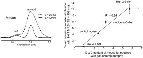
Tissue omega‐3 (ω‐3) content is biologically important to disease; however, its quantification with magnetic resonance spectroscopy in vivo is challenging due to its low concentration. In addition, the ω‐3 methyl resonance (≈ 0.98 ppm) overlaps that of the non‐ω‐3 (≈ 0.90 ppm), even at 9.4 T. We demonstrate that a Point‐RESolved Spectroscopy (PRESS) sequence with an echo time (TE) of 109 ms resolves the ω‐3 and non‐ω‐3 methyl peaks at 9.4 T. Sequence efficacy was verified on five oils with differing ω‐3 fat content; the ω‐3 content obtained correlated with that measured using 16.5 T NMR (R2 = 0.97). The PRESS sequence was also applied to measure ω‐3 content in visceral adipose tissue of three different groups (all n = 3) of mice, each of which were fed a different 20% w/w fat diet. The fat portion of the diet consisted of low (1.4%), medium (9.0%) or high (16.4%) ω‐3 fat. The sequence was also applied to a control mouse fed a standard chow diet (5.6% w/w fat, which was 5.9% ω‐3). Gas chromatography (GC) analysis of excised tissue was performed for each mouse. The ω‐3 fat content obtained with the PRESS sequence correlated with the GC measures (R2 = 0.96). Apparent T2 times of methyl protons were assessed by obtaining spectra from the oils and another group of four mice (fed the high ω‐3 diet) with TE values of 109 and 399 ms. Peak areas were fit to a mono‐exponentially decaying function and the apparent T2 values of the ω‐3 and non‐ω‐3 methyl protons were 906 ± 148 and 398 ± 78 ms, respectively, in the oils. In mice, the values were 410 ± 68 and 283 ± 57 ms for ω‐3 and non‐ω‐3 fats, respectively.
中文翻译:

使用长回波时间磁共振波谱在 9.4 T 下解析小鼠脂肪组织中的 omega-3 甲基共振
组织中的 omega-3 (ω-3) 含量对疾病具有生物学重要性;然而,由于其低浓度,其在体内磁共振波谱法的量化具有挑战性。此外,即使在 9.4 T 时,ω-3 甲基共振(≈ 0.98 ppm)也与非 ω-3 甲基共振(≈ 0.90 ppm)重叠。我们证明了具有回波时间的点分辨光谱(PRESS)序列(TE) 为 109 ms,可分辨 9.4 T 处的 ω-3 和非 ω-3 甲基峰。在具有不同 ω-3 脂肪含量的五种油上验证了序列功效;获得的 ω-3 含量与使用 16.5 T NMR (R 2= 0.97)。PRESS 序列还用于测量三组不同小鼠(所有 n = 3)的内脏脂肪组织中的 ω-3 含量,每组小鼠均饲喂不同的 20% w/w 脂肪饮食。饮食的脂肪部分由低 (1.4%)、中 (9.0%) 或高 (16.4%) ω-3 脂肪组成。该序列还应用于喂食标准食物(5.6% w/w 脂肪,即 5.9% ω-3)的对照小鼠。对每只小鼠进行离体组织的气相色谱 (GC) 分析。用 PRESS 序列获得的 ω-3 脂肪含量与 GC 测量相关(R 2 = 0.96)。表观 T 2通过获得来自油和另一组四只小鼠(喂食高 ω-3 饮食)的光谱来评估甲基质子的时间,TE 值为 109 和 399 ms。峰面积符合单指数衰减函数,油中 ω-3 和非 ω-3 甲基质子的表观 T 2值分别为 906 ± 148 和 398 ± 78 ms。在小鼠中,ω-3 和非 ω-3 脂肪的值分别为 410 ± 68 和 283 ± 57 ms。
更新日期:2021-01-04
中文翻译:
使用长回波时间磁共振波谱在 9.4 T 下解析小鼠脂肪组织中的 omega-3 甲基共振
组织中的 omega-3 (ω-3) 含量对疾病具有生物学重要性;然而,由于其低浓度,其在体内磁共振波谱法的量化具有挑战性。此外,即使在 9.4 T 时,ω-3 甲基共振(≈ 0.98 ppm)也与非 ω-3 甲基共振(≈ 0.90 ppm)重叠。我们证明了具有回波时间的点分辨光谱(PRESS)序列(TE) 为 109 ms,可分辨 9.4 T 处的 ω-3 和非 ω-3 甲基峰。在具有不同 ω-3 脂肪含量的五种油上验证了序列功效;获得的 ω-3 含量与使用 16.5 T NMR (R 2= 0.97)。PRESS 序列还用于测量三组不同小鼠(所有 n = 3)的内脏脂肪组织中的 ω-3 含量,每组小鼠均饲喂不同的 20% w/w 脂肪饮食。饮食的脂肪部分由低 (1.4%)、中 (9.0%) 或高 (16.4%) ω-3 脂肪组成。该序列还应用于喂食标准食物(5.6% w/w 脂肪,即 5.9% ω-3)的对照小鼠。对每只小鼠进行离体组织的气相色谱 (GC) 分析。用 PRESS 序列获得的 ω-3 脂肪含量与 GC 测量相关(R 2 = 0.96)。表观 T 2通过获得来自油和另一组四只小鼠(喂食高 ω-3 饮食)的光谱来评估甲基质子的时间,TE 值为 109 和 399 ms。峰面积符合单指数衰减函数,油中 ω-3 和非 ω-3 甲基质子的表观 T 2值分别为 906 ± 148 和 398 ± 78 ms。在小鼠中,ω-3 和非 ω-3 脂肪的值分别为 410 ± 68 和 283 ± 57 ms。


 京公网安备 11010802027423号