当前位置:
X-MOL 学术
›
ACS Biomater. Sci. Eng.
›
论文详情
Our official English website, www.x-mol.net, welcomes your
feedback! (Note: you will need to create a separate account there.)
Three-Dimensional Bioprinted Hyaluronic Acid Hydrogel Test Beds for Assessing Neural Cell Responses to Competitive Growth Stimuli
ACS Biomaterials Science & Engineering ( IF 5.4 ) Pub Date : 2020-12-01 , DOI: 10.1021/acsbiomaterials.0c00940 Tran B. Ngo 1 , Benjamin S. Spearman 1 , Nora Hlavac 1 , Christine E. Schmidt 1
ACS Biomaterials Science & Engineering ( IF 5.4 ) Pub Date : 2020-12-01 , DOI: 10.1021/acsbiomaterials.0c00940 Tran B. Ngo 1 , Benjamin S. Spearman 1 , Nora Hlavac 1 , Christine E. Schmidt 1
Affiliation
|
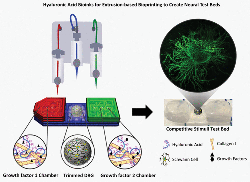
Hyaluronic acid (HA) is an abundant extracellular matrix (ECM) component in soft tissues throughout the body and has found wide adoption in tissue engineering. This study focuses on the optimization of methacrylated HA (MeHA) for three-dimensional (3D) bioprinting to create in vitro test beds that incorporate regeneration-promoting growth factors in neural repair processes. To evaluate MeHA as a potential bioink, rheological studies were performed with PC-12 cells to demonstrate shear thinning properties maintained when printing with and without cells. Next, an extrusion-based Cellink BIO X 3D printer was used to bioprint various MeHA solutions combined with collagen-I to determine which formulation was the most optimal for creating 3D features. Results indicated that MeHA (10 mg/mL) with collagen-I (3 mg/mL) was most suitable. As Schwann cells (SCs) are a critical component of neural repair and regeneration, SC adhesion assessment via integrin β1 immunostaining indicated that the bioink candidate adequately supported SC adhesion and migration when compared to Col-I, a highly cell-adhesive ECM component. MeHA/collagen-I bioink was adapted for neural specific applications by printing with the neural growth factor (NGF) and glial cell line-derived neurotrophic factor (GDNF). These test beds were conducive for SC infiltration and presented differential migration responses. Finally, a two-chamber in vitro test bed design was created to study competitive biochemical cues. Dorsal root ganglia were seeded in test beds and demonstrated directional neurite extension (measured by β-III tubulin and GAP43 immunostaining) in response to NGF and GDNF. Overall, the selected MeHA/collagen-I bioink was bioprintable, improved cell viability compared to molded controls, and was conducive for cell adhesion, growth factor sequestration, and neural cell infiltration. MeHA is a suitable bioink candidate for extrusion-based bioprinting and will be useful in future development of spatially complex test beds to advance in vitro models as an alternative to common in vivo tests for neural repair applications.
中文翻译:

三维生物打印的玻尿酸水凝胶试验床,用于评估神经细胞对竞争性生长刺激的反应。
透明质酸(HA)是人体软组织中丰富的细胞外基质(ECM)成分,已在组织工程中得到广泛采用。这项研究的重点是针对三维(3D)生物打印以创建体外的甲基丙烯酸HA(MeHA)的优化在神经修复过程中结合了促进再生的生长因子的试验床。为了评估MeHA作为潜在的生物墨水,对PC-12细胞进行了流变学研究,以证明在有或没有细胞的情况下打印时均保持剪切稀化特性。接下来,使用基于挤压的Cellink BIO X 3D打印机对结合了胶原蛋白I的各种MeHA解决方案进行生物打印,以确定哪种配方最适合创建3D功能。结果表明,具有胶原蛋白I(3 mg / mL)的MeHA(10 mg / mL)最合适。由于雪旺氏细胞(SCs)是神经修复和再生的关键组成部分,因此通过整合素β1免疫染色表明,与Col-I(一种具有高细胞粘附性的ECM组件)相比,bioink候选药物可充分支持SC粘附和迁移。通过使用神经生长因子(NGF)和神经胶质细胞系衍生的神经营养因子(GDNF)进行印刷,MeHA /胶原蛋白I生物墨水适用于神经特定应用。这些测试床有利于SC渗透,并呈现出不同的迁移响应。最后,进行两室体外实验创建了测试台设计来研究竞争性生化线索。将背根神经节接种在测试床上,并显示对NGF和GDNF的定向神经突延伸(通过β-III微管蛋白和GAP43免疫染色测量)。总体而言,与模制对照相比,所选的MeHA /胶原蛋白I生物墨水可生物打印,可提高细胞活力,并且有利于细胞粘附,生长因子螯合和神经细胞浸润。MeHA是适合基于挤压生物打印的生物墨水候选物,将在空间复杂测试床的未来开发中有用,以推进体外模型,作为神经修复应用中常用的体内测试的替代方法。
更新日期:2020-12-14
中文翻译:
三维生物打印的玻尿酸水凝胶试验床,用于评估神经细胞对竞争性生长刺激的反应。
透明质酸(HA)是人体软组织中丰富的细胞外基质(ECM)成分,已在组织工程中得到广泛采用。这项研究的重点是针对三维(3D)生物打印以创建体外的甲基丙烯酸HA(MeHA)的优化在神经修复过程中结合了促进再生的生长因子的试验床。为了评估MeHA作为潜在的生物墨水,对PC-12细胞进行了流变学研究,以证明在有或没有细胞的情况下打印时均保持剪切稀化特性。接下来,使用基于挤压的Cellink BIO X 3D打印机对结合了胶原蛋白I的各种MeHA解决方案进行生物打印,以确定哪种配方最适合创建3D功能。结果表明,具有胶原蛋白I(3 mg / mL)的MeHA(10 mg / mL)最合适。由于雪旺氏细胞(SCs)是神经修复和再生的关键组成部分,因此通过整合素β1免疫染色表明,与Col-I(一种具有高细胞粘附性的ECM组件)相比,bioink候选药物可充分支持SC粘附和迁移。通过使用神经生长因子(NGF)和神经胶质细胞系衍生的神经营养因子(GDNF)进行印刷,MeHA /胶原蛋白I生物墨水适用于神经特定应用。这些测试床有利于SC渗透,并呈现出不同的迁移响应。最后,进行两室体外实验创建了测试台设计来研究竞争性生化线索。将背根神经节接种在测试床上,并显示对NGF和GDNF的定向神经突延伸(通过β-III微管蛋白和GAP43免疫染色测量)。总体而言,与模制对照相比,所选的MeHA /胶原蛋白I生物墨水可生物打印,可提高细胞活力,并且有利于细胞粘附,生长因子螯合和神经细胞浸润。MeHA是适合基于挤压生物打印的生物墨水候选物,将在空间复杂测试床的未来开发中有用,以推进体外模型,作为神经修复应用中常用的体内测试的替代方法。


 京公网安备 11010802027423号