Chemosphere ( IF 8.1 ) Pub Date : 2020-11-30 , DOI: 10.1016/j.chemosphere.2020.129103 Michela Novelli , Pascale Beffy , Matilde Masini , Chiara Vantaggiato , Luisa Martino , Lorella Marselli , Piero Marchetti , Vincenzo De Tata
|
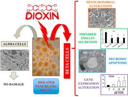
An association between exposure to environmental pollutants and diabetes risk has been repeatedly shown by epidemiological studies. However, the biological basis of this association still need to be clarified. In this research we explored the effects of 2,3,7,8-tetrachlorodibenzo-p-dioxin (TCDD) exposure on isolated pancreatic islets. After 1, 6 and 24 h exposure of isolated islets to different concentrations (1-50 nM) of TCDD we assayed: i) cell survival; ii) ultrastructure; iii) glucose-stimulated insulin secretion (GSIS); iv) expression of selected genes. A significant, dose-related increase of both necrosis and apoptosis was observed isolated rat islets after 24 h exposure to TCDD. The electron microscopic analysis revealed, at the same time point, the presence of several ultrastructural alterations (mitochondrial swelling, increased mitophagy, dilation of the endoplasmic reticulum) that, very interestingly, were exclusively observed in beta cells and not in other endocrine cells. Similar results were obtained in isolated human islets. GSIS was rapidly (1 h) and persistently (6 and 24 h) decreased by TCDD exposure even at the smallest concentration (1 nM). TCDD exposure significantly affected gene expression in isolated islets: Glut2, Gck, Bcl-xL, MafA, Pdx1 FoxO1 and IRE1 gene expression was significantly decreased, whereas Puma, DP5, iNOS and Chop gene expression was significantly increased after 6 h exposure to TCDD. In conclusion, our results clearly indicated that pancreatic beta cells represent not only a sensitive but also a specific target of the toxic action of dioxin.
中文翻译:
2,3,7,8-四氯二苯并偶氮对-二恶英在分离的胰岛上的选择性贝塔细胞毒性
流行病学研究已反复表明暴露于环境污染物与糖尿病风险之间的关联。但是,这种关联的生物学基础仍然需要阐明。在这项研究中,我们探讨了2,3,7,8-四氯二苯并-对-二恶英(TCDD)暴露对孤立的胰岛的影响。在将分离的胰岛暴露于不同浓度(1-50 nM)的TCDD 1、6和24小时后,我们进行了以下测定:i)细胞存活;ii)超微结构;iii)葡萄糖刺激的胰岛素分泌(GSIS);iv)所选基因的表达。暴露于TCDD 24小时后,分离的大鼠胰岛观察到坏死和凋亡的剂量相关显着增加。电镜分析显示,在同一时间点,存在几种超微结构改变(线粒体肿胀,线粒体增多,非常有趣的是,仅在内膜细胞中观察到内质网的扩张),而在其他内分泌细胞中未观察到。在分离的人胰岛中获得了相似的结果。即使在最小浓度(1 nM)下,TCDD暴露也会使GSIS迅速(1 h)持续下降(6和24 h)。TCDD暴露显着影响离体胰岛的基因表达:Glut2,Gck,Bcl-xL,MafA,Pdx1 FoxO1和IRE1基因表达显着降低,而Puma,DP5,iNOS和Chop基因表达在TCDD暴露6 h后显着增加。总之,我们的结果清楚地表明,胰腺β细胞不仅代表了二恶英毒性作用的敏感目标,而且还代表了其特定的靶标。仅在β细胞中观察到,而在其他内分泌细胞中观察不到。在分离的人胰岛中获得了相似的结果。即使在最小浓度(1 nM)下,TCDD暴露也会使GSIS迅速(1 h)持续下降(6和24 h)。TCDD暴露显着影响离体胰岛的基因表达:Glut2,Gck,Bcl-xL,MafA,Pdx1 FoxO1和IRE1基因表达显着降低,而Puma,DP5,iNOS和Chop基因表达在TCDD暴露6 h后显着增加。总之,我们的结果清楚地表明,胰腺β细胞不仅代表了二恶英毒性作用的敏感目标,而且还代表了其特定的靶标。仅在β细胞中观察到,而在其他内分泌细胞中观察不到。在分离的人胰岛中获得了相似的结果。即使在最小浓度(1 nM)下,TCDD暴露也会使GSIS迅速(1 h)持续下降(6和24 h)。TCDD暴露显着影响离体胰岛的基因表达:Glut2,Gck,Bcl-xL,MafA,Pdx1 FoxO1和IRE1基因表达显着降低,而Puma,DP5,iNOS和Chop基因表达在TCDD暴露6 h后显着增加。总之,我们的结果清楚地表明,胰腺β细胞不仅代表了二恶英毒性作用的敏感目标,而且还代表了其特定的靶标。即使在最小浓度(1 nM)下,TCDD暴露也会使GSIS迅速(1 h)持续下降(6和24 h)。TCDD暴露显着影响离体胰岛的基因表达:Glut2,Gck,Bcl-xL,MafA,Pdx1 FoxO1和IRE1基因表达显着降低,而Puma,DP5,iNOS和Chop基因表达在TCDD暴露6 h后显着增加。总之,我们的结果清楚地表明,胰腺β细胞不仅代表了二恶英毒性作用的敏感目标,而且还代表了其特定的靶标。即使在最小浓度(1 nM)下,TCDD暴露也会使GSIS迅速(1 h)持续下降(6和24 h)。TCDD暴露显着影响离体胰岛的基因表达:Glut2,Gck,Bcl-xL,MafA,Pdx1 FoxO1和IRE1基因表达显着降低,而Puma,DP5,iNOS和Chop基因表达在TCDD暴露6 h后显着增加。总之,我们的结果清楚地表明,胰腺β细胞不仅代表了二恶英毒性作用的敏感目标,而且还代表了其特定的靶标。


 京公网安备 11010802027423号