当前位置:
X-MOL 学术
›
Inorg. Chem. Commun.
›
论文详情
Our official English website, www.x-mol.net, welcomes your
feedback! (Note: you will need to create a separate account there.)
Disaccharide assisted LaAlO3:Ce3+ perovskite: structural and optical studies suitable for display devices
Inorganic Chemistry Communications ( IF 4.4 ) Pub Date : 2021-01-01 , DOI: 10.1016/j.inoche.2020.108342 Yashaswini , S. Pratibha , R. Lokesh , N. Dhananjaya , C. Pandurangappa
Inorganic Chemistry Communications ( IF 4.4 ) Pub Date : 2021-01-01 , DOI: 10.1016/j.inoche.2020.108342 Yashaswini , S. Pratibha , R. Lokesh , N. Dhananjaya , C. Pandurangappa
|
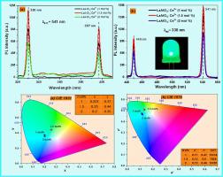
Abstract Sucrose mediated LaAlO3: Ce3+ nanopowders were prepared by propellant chemistry technique. The preliminary characterizations were carried out using Powder X-Ray Diffraction (PXRD), Fourier Transform InfraRed spectra (FTIR), Scanning Electron Microscopy (SEM) and transmission electron microscopy (TEM) to analyse the structure and morphology of the prepared nanophosphors. PXRD studies showed pure rhombohedral single phase with average crystallite size of the order of 45-62 nm. SEM micrographs confirmed nanoparticles formation and the crystallite size agrees well with the TEM image. The optical band gap of LaAlO3: Ce3+ (1-2 mol%) nanophosphors was found to be in the range 4.5-4.93 eV. PL spectra showed broad intense band at 449 nm and 541 nm up on 330 nm excitation. Chromaticity studies exhibit the blue green color emission of the prepared phosphors. The intensity and color of Ce3+ doped LaAlO3 nanophosphors confirm that these phosphors can be a potential material for display and optical devices.
中文翻译:

双糖辅助 LaAlO3:Ce3+ 钙钛矿:适用于显示设备的结构和光学研究
摘要 采用推进剂化学技术制备了蔗糖介导的LaAlO3:Ce3+纳米粉体。使用粉末 X 射线衍射 (PXRD)、傅里叶变换红外光谱 (FTIR)、扫描电子显微镜 (SEM) 和透射电子显微镜 (TEM) 进行初步表征,以分析制备的纳米磷光体的结构和形貌。PXRD 研究显示纯菱面体单相,平均晶粒尺寸为 45-62 nm。SEM 显微照片证实了纳米颗粒的形成,并且微晶尺寸与 TEM 图像非常吻合。发现 LaAlO3: Ce3+ (1-2 mol%) 纳米磷光体的光学带隙在 4.5-4.93 eV 范围内。PL 光谱在 449 nm 和 541 nm 处在 330 nm 激发处显示出宽的强谱带。色度研究显示所制备的磷光体的蓝绿色发射。Ce3+ 掺杂的 LaAlO3 纳米磷光体的强度和颜色证实,这些磷光体可以成为显示和光学设备的潜在材料。
更新日期:2021-01-01
中文翻译:
双糖辅助 LaAlO3:Ce3+ 钙钛矿:适用于显示设备的结构和光学研究
摘要 采用推进剂化学技术制备了蔗糖介导的LaAlO3:Ce3+纳米粉体。使用粉末 X 射线衍射 (PXRD)、傅里叶变换红外光谱 (FTIR)、扫描电子显微镜 (SEM) 和透射电子显微镜 (TEM) 进行初步表征,以分析制备的纳米磷光体的结构和形貌。PXRD 研究显示纯菱面体单相,平均晶粒尺寸为 45-62 nm。SEM 显微照片证实了纳米颗粒的形成,并且微晶尺寸与 TEM 图像非常吻合。发现 LaAlO3: Ce3+ (1-2 mol%) 纳米磷光体的光学带隙在 4.5-4.93 eV 范围内。PL 光谱在 449 nm 和 541 nm 处在 330 nm 激发处显示出宽的强谱带。色度研究显示所制备的磷光体的蓝绿色发射。Ce3+ 掺杂的 LaAlO3 纳米磷光体的强度和颜色证实,这些磷光体可以成为显示和光学设备的潜在材料。


 京公网安备 11010802027423号