当前位置:
X-MOL 学术
›
ACS Biomater. Sci. Eng.
›
论文详情
Our official English website, www.x-mol.net, welcomes your
feedback! (Note: you will need to create a separate account there.)
Rare-Earth-Doped Cerium Oxide Nanocubes for Biomedical Near-Infrared and Magnetic Resonance Imaging
ACS Biomaterials Science & Engineering ( IF 5.4 ) Pub Date : 2020-11-24 , DOI: 10.1021/acsbiomaterials.0c01193 Anne E D'Achille 1 , Roberto Gonzalez-Rodriguez 1 , Elizabeth Campbell 2 , Bong Han Lee 2 , Jeffery L Coffer 1 , Anton V Naumov 2
ACS Biomaterials Science & Engineering ( IF 5.4 ) Pub Date : 2020-11-24 , DOI: 10.1021/acsbiomaterials.0c01193 Anne E D'Achille 1 , Roberto Gonzalez-Rodriguez 1 , Elizabeth Campbell 2 , Bong Han Lee 2 , Jeffery L Coffer 1 , Anton V Naumov 2
Affiliation
|
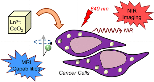
Near-infrared (NIR) fluorescence provides a new avenue for biomedical fluorescence imaging that allows for the tracking of fluorophore through several centimeters of biological tissue. However, such fluorophores are rare and, due to accumulation-derived toxicity, are often restricted from clinical applications. Deep tissue imaging not only provided by near-infrared fluorophores but also conventionally carried out by magnetic resonance imaging (MRI) or computed tomography (CT) is also hampered by the toxicity of the contrast agents. This work offers a biocompatible imaging solution: cerium oxide (CeO2) nanocubes doped with ytterbium or neodymium, and co-doped with gadolinium, showing simultaneous potential for near-infrared (NIR) fluorescence and magnetic resonance imaging (MRI) applications. A synthetic process described in this work allows for the stable incorporation of ytterbium or neodymium, both possessing emissive transitions in the NIR. As a biocompatible nanomaterial, the CeO2 nanocubes act as an ideal host material for doping, minimizing lanthanide fluorescence self-quenching as well as any potential toxicity associated with the dopants. The uptake of nanocubes by HeLa cells maximized at 12 h was monitored by hyperspectral imaging of the ytterbium or neodymium NIR emission, indicating the capacity of the lanthanide-doped nanocubes for in vitro and a potential for in vivo fluorescence imaging. The co-doped nanocubes demonstrate no significant loss of NIR emission intensity upon co-doping with 2 atomic % gadolinium and exhibit magnetic susceptibilities in the range of known negative contrast agents. However, a small increase to 6 atomic % gadolinium significantly affects the magnetic susceptibility ratio (r2/r1), shifting closer to the positive contrast range and suggesting the potential use of the CeO2 nanocube matrix doped with selected rare-earth ions as a tunable MRI contrast agent with NIR imaging capabilities.
中文翻译:

用于生物医学近红外和磁共振成像的稀土掺杂氧化铈纳米立方体
近红外 (NIR) 荧光为生物医学荧光成像提供了一条新途径,允许通过几厘米的生物组织跟踪荧光团。然而,这种荧光团很少见,并且由于积累衍生的毒性,通常限制临床应用。深部组织成像不仅由近红外荧光团提供,而且通常由磁共振成像 (MRI) 或计算机断层扫描 (CT) 进行,也受到造影剂毒性的阻碍。这项工作提供了一种生物相容性成像解决方案:氧化铈(CeO 2) 掺杂镱或钕以及共掺杂钆的纳米立方体,显示出同时用于近红外 (NIR) 荧光和磁共振成像 (MRI) 应用的潜力。这项工作中描述的合成过程允许稳定掺入镱或钕,两者都在 NIR 中具有发射跃迁。作为一种生物相容性纳米材料,CeO 2纳米立方体可作为一种理想的掺杂主体材料,最大限度地减少镧系元素荧光自猝灭以及与掺杂剂相关的任何潜在毒性。通过镱或钕 NIR 发射的高光谱成像监测 HeLa 细胞在 12 小时时对纳米立方体的最大吸收,表明镧系元素掺杂纳米立方体的体外容量和潜在的体内荧光成像。共掺杂的纳米立方体在与 2 原子 % 的钆共掺杂时没有显示出 NIR 发射强度的显着损失,并且表现出在已知负造影剂范围内的磁化率。然而,钆的小幅增加至 6 原子 % 会显着影响磁化率比 ( r 2 / r 1 ),移动到更接近正对比度范围,并表明掺杂有选定稀土离子的 CeO 2纳米立方体基质的潜在用途作为具有 NIR 成像能力的可调 MRI 造影剂。
更新日期:2020-12-14
中文翻译:
用于生物医学近红外和磁共振成像的稀土掺杂氧化铈纳米立方体
近红外 (NIR) 荧光为生物医学荧光成像提供了一条新途径,允许通过几厘米的生物组织跟踪荧光团。然而,这种荧光团很少见,并且由于积累衍生的毒性,通常限制临床应用。深部组织成像不仅由近红外荧光团提供,而且通常由磁共振成像 (MRI) 或计算机断层扫描 (CT) 进行,也受到造影剂毒性的阻碍。这项工作提供了一种生物相容性成像解决方案:氧化铈(CeO 2) 掺杂镱或钕以及共掺杂钆的纳米立方体,显示出同时用于近红外 (NIR) 荧光和磁共振成像 (MRI) 应用的潜力。这项工作中描述的合成过程允许稳定掺入镱或钕,两者都在 NIR 中具有发射跃迁。作为一种生物相容性纳米材料,CeO 2纳米立方体可作为一种理想的掺杂主体材料,最大限度地减少镧系元素荧光自猝灭以及与掺杂剂相关的任何潜在毒性。通过镱或钕 NIR 发射的高光谱成像监测 HeLa 细胞在 12 小时时对纳米立方体的最大吸收,表明镧系元素掺杂纳米立方体的体外容量和潜在的体内荧光成像。共掺杂的纳米立方体在与 2 原子 % 的钆共掺杂时没有显示出 NIR 发射强度的显着损失,并且表现出在已知负造影剂范围内的磁化率。然而,钆的小幅增加至 6 原子 % 会显着影响磁化率比 ( r 2 / r 1 ),移动到更接近正对比度范围,并表明掺杂有选定稀土离子的 CeO 2纳米立方体基质的潜在用途作为具有 NIR 成像能力的可调 MRI 造影剂。


 京公网安备 11010802027423号