当前位置:
X-MOL 学术
›
NMR Biomed.
›
论文详情
Our official English website, www.x-mol.net, welcomes your feedback! (Note: you will need to create a separate account there.)
Identification of variable stages in murine pancreatic tumors by a multiparametric approach employing hyperpolarized 13C MRSI, 1H diffusivity and 1H T1 MRI
NMR in Biomedicine ( IF 2.9 ) Pub Date : 2020-11-21 , DOI: 10.1002/nbm.4446 Ricardo P Martinho 1 , Qingjia Bao 1 , Stefan Markovic 1 , Dina Preise 2 , Keren Sasson 2 , Lilach Agemy 3 , Avigdor Scherz 3 , Lucio Frydman 1
NMR in Biomedicine ( IF 2.9 ) Pub Date : 2020-11-21 , DOI: 10.1002/nbm.4446 Ricardo P Martinho 1 , Qingjia Bao 1 , Stefan Markovic 1 , Dina Preise 2 , Keren Sasson 2 , Lilach Agemy 3 , Avigdor Scherz 3 , Lucio Frydman 1
Affiliation
|
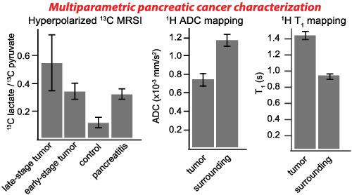
This study explored the usefulness of multiple quantitative MRI approaches to detect pancreatic ductal adenocarcinomas in two murine models, PAN‐02 and KPC. Methods assayed included 1H T1 and T2 measurements, quantitative diffusivity mapping, magnetization transfer (MT) 1H MRI throughout the abdomen and hyperpolarized 13C spectroscopic imaging. The progress of the disease was followed as a function of its development; studies were also conducted for wildtype control mice and for mice with induced mild acute pancreatitis. Customized methods developed for scanning the motion‐ and artifact‐prone mice abdomens allowed us to obtain quality 1H images for these targeted regions. Contrasts between tumors and surrounding tissues, however, were significantly different. Anatomical images, T2 maps and MT did not yield significant contrast unless tumors were large. By contrast, tumors showed statistically lower diffusivities than their surroundings (≈8.3 ± 0.4 x 10⁻⁴ for PAN‐02 and ≈10.2 ± 0.6 x 10⁻⁴ for KPC vs 13 ± 1 x 10⁻³ mm2 s−1 for surroundings), longer T1 relaxation times (≈1.44 ± 0.05 for PAN‐02 and ≈1.45 ± 0.05 for KPC vs 0.95 ± 0.10 seconds for surroundings) and significantly higher lactate/pyruvate ratios by hyperpolarized 13C MR (0.53 ± 0.2 for PAN‐02 and 0.78 ± 0.2 for KPC vs 0.11 ± 0.04 for control and 0.31 ± 0.04 for pancreatitis‐bearing mice). Although the latter could also distinguish early‐stage tumors from healthy animal controls, their response was similar to that in our pancreatitis model. Still, this ambiguity could be lifted using the 1H‐based reporters. If confirmed for other kinds of pancreatic tumors this means that these approaches, combined, can provide a route to an early detection of pancreatic cancer.
中文翻译:

通过采用超极化 13C MRSI、1H 扩散率和 1H T1 MRI 的多参数方法识别小鼠胰腺肿瘤的不同阶段
本研究探讨了多种定量 MRI 方法在两种小鼠模型 PAN-02 和 KPC 中检测胰腺导管腺癌的有效性。测定的方法包括1 H T 1和 T 2测量、定量扩散映射、整个腹部的磁化转移 (MT) 1 H MRI 和超极化13 C 光谱成像。疾病的进展作为其发展的函数被跟踪;还对野生型对照小鼠和诱发轻度急性胰腺炎的小鼠进行了研究。为扫描易发生运动和伪影的小鼠腹部而开发的定制方法使我们能够获得质量1这些目标区域的 H 图像。然而,肿瘤与周围组织之间的对比显着不同。除非肿瘤很大,否则解剖图像、T 2图和 MT 不会产生显着的对比度。相比之下,肿瘤显示出统计学上较低的扩散率比其周围(≈8.3±0.4×10 -4对PAN-02和≈10.2±0.6×10 -4对KPC对13±1×10 -3毫米2个小号-1为环境),更长的 T 1弛豫时间(PAN-02 ≈1.44 ± 0.05,KPC ≈1.45 ± 0.05,周围环境为 0.95 ± 0.10 秒)和超极化13显着更高的乳酸/丙酮酸比率C MR(PAN-02 为 0.53 ± 0.2,KPC 为 0.78 ± 0.2,对照为 0.11 ± 0.04,胰腺炎小鼠为 0.31 ± 0.04)。尽管后者也可以将早期肿瘤与健康动物对照区分开来,但它们的反应与我们的胰腺炎模型中的反应相似。尽管如此,使用基于1 H 的记者可以消除这种歧义。如果证实其他类型的胰腺肿瘤,这意味着这些方法结合起来,可以提供一条早期检测胰腺癌的途径。
更新日期:2021-01-04
中文翻译:
通过采用超极化 13C MRSI、1H 扩散率和 1H T1 MRI 的多参数方法识别小鼠胰腺肿瘤的不同阶段
本研究探讨了多种定量 MRI 方法在两种小鼠模型 PAN-02 和 KPC 中检测胰腺导管腺癌的有效性。测定的方法包括1 H T 1和 T 2测量、定量扩散映射、整个腹部的磁化转移 (MT) 1 H MRI 和超极化13 C 光谱成像。疾病的进展作为其发展的函数被跟踪;还对野生型对照小鼠和诱发轻度急性胰腺炎的小鼠进行了研究。为扫描易发生运动和伪影的小鼠腹部而开发的定制方法使我们能够获得质量1这些目标区域的 H 图像。然而,肿瘤与周围组织之间的对比显着不同。除非肿瘤很大,否则解剖图像、T 2图和 MT 不会产生显着的对比度。相比之下,肿瘤显示出统计学上较低的扩散率比其周围(≈8.3±0.4×10 -4对PAN-02和≈10.2±0.6×10 -4对KPC对13±1×10 -3毫米2个小号-1为环境),更长的 T 1弛豫时间(PAN-02 ≈1.44 ± 0.05,KPC ≈1.45 ± 0.05,周围环境为 0.95 ± 0.10 秒)和超极化13显着更高的乳酸/丙酮酸比率C MR(PAN-02 为 0.53 ± 0.2,KPC 为 0.78 ± 0.2,对照为 0.11 ± 0.04,胰腺炎小鼠为 0.31 ± 0.04)。尽管后者也可以将早期肿瘤与健康动物对照区分开来,但它们的反应与我们的胰腺炎模型中的反应相似。尽管如此,使用基于1 H 的记者可以消除这种歧义。如果证实其他类型的胰腺肿瘤,这意味着这些方法结合起来,可以提供一条早期检测胰腺癌的途径。


 京公网安备 11010802027423号