Our official English website, www.x-mol.net, welcomes your
feedback! (Note: you will need to create a separate account there.)
Myeloid‐derived suppressor cells support remyelination in a murine model of multiple sclerosis by promoting oligodendrocyte precursor cell survival, proliferation, and differentiation
Glia ( IF 5.4 ) Pub Date : 2020-11-20 , DOI: 10.1002/glia.23936 Carolina Melero-Jerez 1, 2 , Beatriz Fernández-Gómez 1 , Rafael Lebrón-Galán 2 , Maria Cristina Ortega 2 , Irene Sánchez-de Lara 2 , Ana Cristina Ojalvo 2 , Diego Clemente 2 , Fernando de Castro 1
Glia ( IF 5.4 ) Pub Date : 2020-11-20 , DOI: 10.1002/glia.23936 Carolina Melero-Jerez 1, 2 , Beatriz Fernández-Gómez 1 , Rafael Lebrón-Galán 2 , Maria Cristina Ortega 2 , Irene Sánchez-de Lara 2 , Ana Cristina Ojalvo 2 , Diego Clemente 2 , Fernando de Castro 1
Affiliation
|
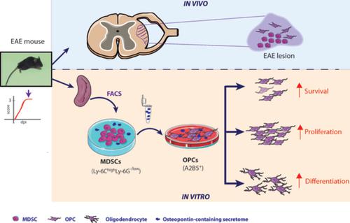
The most frequent variant of multiple sclerosis (MS) is the relapsing–remitting form, characterized by symptomatic phases followed by periods of total/partial recovery. Hence, it is possible that these patients can benefit from endogenous agents that control the inflammatory process and favor spontaneous remyelination. In this context, there is increasing interest in the role of myeloid‐derived suppressor cells (MDSCs) during the clinical course of experimental autoimmune encephalomyelitis (EAE). MDSCs speed up infiltrated T‐cell anergy and apoptosis. In different animal models of MS, a milder disease course is related to higher presence/density of MDSCs in the periphery, and smaller demyelinated lesions in the central nervous system (CNS). These observations lead us to wonder whether MDSCs might not only exert an anti‐inflammatory effect but might also have direct influence on oligodendrocyte precursor cells (OPCs) and remyelination. In the present work, we reveal for the first time the relationship between OPCs and MDSCs in EAE, relationship that is guided by the distance from the inflammatory core. We describe the effects of MDSCs on survival, proliferation, as well as potent promoters of OPC differentiation toward mature phenotypes. We show for the first time that osteopontin is remarkably present in the analyzed secretome of MDSCs. The ablation of this cue from MDSCs‐secretome demonstrates that osteopontin is the main MDSC effector on these oligodendroglial cells. These data highlight a crucial pathogenic interaction between innate immunity and the CNS, opening ways to develop MDSC‐ and/or osteopontin‐based therapies to promote effective myelin preservation and repair in MS patients.
中文翻译:

髓源性抑制细胞通过促进少突胶质前体细胞存活、增殖和分化来支持多发性硬化小鼠模型中的髓鞘再生
多发性硬化症 (MS) 最常见的变体是复发-缓解形式,其特征在于症状阶段,然后是完全/部分恢复期。因此,这些患者可能会受益于控制炎症过程并有利于自发髓鞘再生的内源性药物。在这种情况下,人们对髓源性抑制细胞(MDSCs)在实验性自身免疫性脑脊髓炎(EAE)临床过程中的作用越来越感兴趣。MDSC 加速浸润的 T 细胞无反应和凋亡。在 MS 的不同动物模型中,较轻的病程与外周 MDSC 的存在/密度较高以及中枢神经系统 (CNS) 中较小的脱髓鞘病变有关。这些观察让我们想知道 MDSCs 是否不仅可能发挥抗炎作用,而且可能对少突胶质前体细胞 (OPCs) 和髓鞘再生有直接影响。在目前的工作中,我们首次揭示了 EAE 中 OPCs 和 MDSCs 之间的关系,这种关系由与炎症核心的距离引导。我们描述了 MDSC 对存活、增殖的影响,以及 OPC 向成熟表型分化的有效促进剂。我们首次表明骨桥蛋白显着存在于分析的 MDSC 分泌组中。从 MDSCs-secretome 消除这一线索表明,骨桥蛋白是这些少突胶质细胞上的主要 MDSC 效应物。这些数据突出了先天免疫和中枢神经系统之间至关重要的致病相互作用,
更新日期:2020-11-20
中文翻译:
髓源性抑制细胞通过促进少突胶质前体细胞存活、增殖和分化来支持多发性硬化小鼠模型中的髓鞘再生
多发性硬化症 (MS) 最常见的变体是复发-缓解形式,其特征在于症状阶段,然后是完全/部分恢复期。因此,这些患者可能会受益于控制炎症过程并有利于自发髓鞘再生的内源性药物。在这种情况下,人们对髓源性抑制细胞(MDSCs)在实验性自身免疫性脑脊髓炎(EAE)临床过程中的作用越来越感兴趣。MDSC 加速浸润的 T 细胞无反应和凋亡。在 MS 的不同动物模型中,较轻的病程与外周 MDSC 的存在/密度较高以及中枢神经系统 (CNS) 中较小的脱髓鞘病变有关。这些观察让我们想知道 MDSCs 是否不仅可能发挥抗炎作用,而且可能对少突胶质前体细胞 (OPCs) 和髓鞘再生有直接影响。在目前的工作中,我们首次揭示了 EAE 中 OPCs 和 MDSCs 之间的关系,这种关系由与炎症核心的距离引导。我们描述了 MDSC 对存活、增殖的影响,以及 OPC 向成熟表型分化的有效促进剂。我们首次表明骨桥蛋白显着存在于分析的 MDSC 分泌组中。从 MDSCs-secretome 消除这一线索表明,骨桥蛋白是这些少突胶质细胞上的主要 MDSC 效应物。这些数据突出了先天免疫和中枢神经系统之间至关重要的致病相互作用,


 京公网安备 11010802027423号