当前位置:
X-MOL 学术
›
NeuroImage
›
论文详情
Our official English website, www.x-mol.net, welcomes your
feedback! (Note: you will need to create a separate account there.)
Multimodal 3D atlas of the macaque monkey motor and premotor cortex
NeuroImage ( IF 4.7 ) Pub Date : 2021-02-01 , DOI: 10.1016/j.neuroimage.2020.117574 Lucija Rapan 1 , Sean Froudist-Walsh 2 , Meiqi Niu 1 , Ting Xu 3 , Thomas Funck 1 , Karl Zilles 1 , Nicola Palomero-Gallagher 4
NeuroImage ( IF 4.7 ) Pub Date : 2021-02-01 , DOI: 10.1016/j.neuroimage.2020.117574 Lucija Rapan 1 , Sean Froudist-Walsh 2 , Meiqi Niu 1 , Ting Xu 3 , Thomas Funck 1 , Karl Zilles 1 , Nicola Palomero-Gallagher 4
Affiliation
|
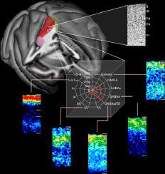
In the present study we reevaluated the parcellation scheme of the macaque frontal agranular cortex by implementing quantitative cytoarchitectonic and multireceptor analyses, with the purpose to integrate and reconcile the discrepancies between previously published maps of this region. We applied an observer-independent and statistically testable approach to determine the position of cytoarchitectonic borders. Analysis of the regional and laminar distribution patterns of 13 different transmitter receptors confirmed the position of cytoarchitectonically identified borders. Receptor densities were extracted from each area and visualized as its "receptor fingerprint". Hierarchical and principal components analyses were conducted to detect clusters of areas according to the degree of (dis)similarity of their fingerprints. Finally, functional connectivity pattern of each identified area was analyzed with areas of prefrontal, cingulate, somatosensory and lateral parietal cortex and the results were depicted as "connectivity fingerprints" and seed-to-vertex connectivity maps. We identified 16 cyto- and receptor architectonically distinct areas, including novel subdivisions of the primary motor area 4 (i.e. 4a, 4p, 4m) and of premotor areas F4 (i.e. F4s, F4d, F4v), F5 (i.e. F5s, F5d, F5v) and F7 (i.e. F7d, F7i, F7s). Multivariate analyses of receptor fingerprints revealed three clusters, which first segregated the subdivisions of area 4 with F4d and F4s from the remaining premotor areas, then separated ventrolateral from dorsolateral and medial premotor areas. The functional connectivity analysis revealed that medial and dorsolateral premotor and motor areas show stronger functional connectivity with areas involved in visual processing, whereas 4p and ventrolateral premotor areas presented a stronger functional connectivity with areas involved in somatomotor responses. For the first time, we provide a 3D atlas integrating cyto- and multi-receptor architectonic features of the macaque motor and premotor cortex. This atlas constitutes a valuable resource for the analysis of functional experiments carried out with non-human primates, for modeling approaches with realistic synaptic dynamics, as well as to provide insights into how brain functions have developed by changes in the underlying microstructure and encoding strategies during evolution.
中文翻译:

猕猴运动和前运动皮层的多模态 3D 图谱
在本研究中,我们通过实施定量细胞结构和多受体分析重新评估了猕猴额叶无颗粒皮层的分区方案,目的是整合和协调先前发布的该区域地图之间的差异。我们采用独立于观察者且可统计测试的方法来确定细胞结构边界的位置。对 13 种不同递质受体的区域和层状分布模式的分析证实了细胞结构上识别的边界的位置。从每个区域提取受体密度并可视化为其“受体指纹”。进行层次分析和主成分分析,根据指纹的相似(不)相似程度来检测区域的聚类。最后,对前额叶、扣带皮层、体感和外侧顶叶皮层区域的每个识别区域的功能连接模式进行了分析,并将结果描述为“连接指纹”和种子到顶点的连接图。我们鉴定了 16 个细胞和受体结构上不同的区域,包括初级运动区 4(即 4a、4p、4m)和前运动区 F4(即 F4s、F4d、F4v)、F5(即 F5s、F5d、F5v)的新细分。 )和F7(即F7d、F7i、F7s)。受体指纹的多变量分析揭示了三个簇,首先将具有 F4d 和 F4s 的区域 4 的细分与其余运动前区域分开,然后将腹外侧与背外侧和内侧运动前区域分开。 功能连接分析显示,内侧和背外侧运动前区和运动区与涉及视觉处理的区域表现出更强的功能连接,而4p和腹外侧运动前区与涉及躯体运动反应的区域表现出更强的功能连接。我们首次提供了整合猕猴运动和前运动皮层的细胞和多受体结构特征的 3D 图谱。该图谱为分析非人类灵长类动物的功能实验、具有真实突触动力学的建模方法以及提供关于大脑功能如何通过潜在微观结构和编码策略的变化而发展的见解提供了宝贵的资源。进化。
更新日期:2021-02-01
中文翻译:
猕猴运动和前运动皮层的多模态 3D 图谱
在本研究中,我们通过实施定量细胞结构和多受体分析重新评估了猕猴额叶无颗粒皮层的分区方案,目的是整合和协调先前发布的该区域地图之间的差异。我们采用独立于观察者且可统计测试的方法来确定细胞结构边界的位置。对 13 种不同递质受体的区域和层状分布模式的分析证实了细胞结构上识别的边界的位置。从每个区域提取受体密度并可视化为其“受体指纹”。进行层次分析和主成分分析,根据指纹的相似(不)相似程度来检测区域的聚类。最后,对前额叶、扣带皮层、体感和外侧顶叶皮层区域的每个识别区域的功能连接模式进行了分析,并将结果描述为“连接指纹”和种子到顶点的连接图。我们鉴定了 16 个细胞和受体结构上不同的区域,包括初级运动区 4(即 4a、4p、4m)和前运动区 F4(即 F4s、F4d、F4v)、F5(即 F5s、F5d、F5v)的新细分。 )和F7(即F7d、F7i、F7s)。受体指纹的多变量分析揭示了三个簇,首先将具有 F4d 和 F4s 的区域 4 的细分与其余运动前区域分开,然后将腹外侧与背外侧和内侧运动前区域分开。 功能连接分析显示,内侧和背外侧运动前区和运动区与涉及视觉处理的区域表现出更强的功能连接,而4p和腹外侧运动前区与涉及躯体运动反应的区域表现出更强的功能连接。我们首次提供了整合猕猴运动和前运动皮层的细胞和多受体结构特征的 3D 图谱。该图谱为分析非人类灵长类动物的功能实验、具有真实突触动力学的建模方法以及提供关于大脑功能如何通过潜在微观结构和编码策略的变化而发展的见解提供了宝贵的资源。进化。


 京公网安备 11010802027423号