Biomaterials Advances ( IF 5.5 ) Pub Date : 2020-11-21 , DOI: 10.1016/j.msec.2020.111739 Akram Shafiee , Mousa Kehtari , Zeinab Zarei , Masoud Soleimani , Reyhaneh Varshochian , Amirhossein Ahmadi , Fatemeh Atyabi , Rassoul Dinarvand
|
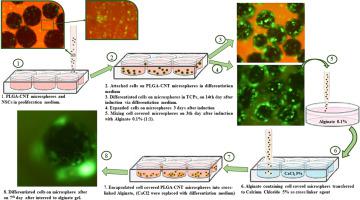
The cell-extracellular matrix (ECM) interactions are known to have a strong impact on cell behaviors in neural tissues. Due to complex physiology system and limited regenerative capacity of nervous system, neural tissue engineering has attracted attention as a promising strategy. In this study, we designed a hydrogel loaded by poly (lactic-co-glycolic acid) (PLGA) microspheres containing carbon nanotubes (CNT) and the biochemical differentiation factors, as a scaffold, in order to replicate the neural niche for stem cell growth (and/or differentiation). Different formulations from Hyaluronic acid (H), Poloxamer (P), Ethoxy-silane-capped poloxamer (PE), and cross-linked Alginate (Alg) were utilized as an in situ gel structure matrix to mirror the mechanical properties of the ECM of CNS. Subsequently, conductivity, surface morphology, size of microspheres, and CNT dispersion in microsphere were measured using two probes electrical conductometer, scanning electron microscopy (SEM), dynamic light scattering (DLS), and Raman spectroscopy, respectively. According to SEM and fluorescent microscopy images, CNTs increased the porosity of polymeric structure, which, in turn, facilitated the adhesion of stem cells on the surface of microspheres compared with control. Microstructure and rheological behaviors of different gel compositions were investigated using SEM and parallel-plate oscillatory rheometer, respectively. The MTT assay showed the toxicity profile of hydrogels was appropriate for cell transplantation. The confocal images illustrated the 3D platform of P15%H10% and P20%H5% gel formulations containing the PLGA-CNT microspheres, which allows the proliferation of neural stem cells (NSCs) derived from MSC. The results of real-time PCR and immunocytochemistry showed neuronal differentiation capacity of cultured NSCs derived from MSC in the alginate gel that contained PLGA-CNT microspheres as well as other control groups. The dispersion of the CNT-PLGA microspheres, covered by NSCs, into alginate gel in the presence of induction factors was found to notably enhance the expression of Sox2-SYP and β-Tubulin III neuronal markers.
中文翻译:
PLGA微球负载的原位形成水凝胶的支架,其中包含碳纳米管作为神经分化的合适位
已知细胞与细胞外基质(ECM)的相互作用对神经组织中的细胞行为有很大影响。由于复杂的生理系统和神经系统的再生能力有限,神经组织工程作为一种有前途的策略引起了人们的关注。在这项研究中,我们旨在通过聚(乳酸-装水凝胶合作-乙醇酸(PLGA)微球包含碳纳米管(CNT)和生化分化因子,作为支架,以便复制干细胞生长(和/或分化)的神经环境。透明质酸(H),泊洛沙姆(P),乙氧基硅烷封端的泊洛沙姆(PE)和交联的藻酸盐(Alg)的不同配方被用作原位凝胶结构基质,以反映ECM的ECM的机械性能。 CNS。随后,分别使用两个探针电导仪,扫描电子显微镜(SEM),动态光散射(DLS)和拉曼光谱仪测量电导率,表面形态,微球尺寸和CNT在微球中的分散度。根据SEM和荧光显微镜图像,碳纳米管增加了聚合物结构的孔隙率,进而,与对照相比,促进干细胞在微球表面的粘附。分别使用SEM和平行板振荡流变仪研究了不同凝胶组成的微观结构和流变行为。MTT分析表明水凝胶的毒性特征适合于细胞移植。共聚焦图像说明了含有PLGA-CNT微球的P15%H10%和P20%H5%凝胶制剂的3D平台,该平台允许衍生自MSC的神经干细胞(NSC)增殖。实时PCR和免疫细胞化学的结果表明,在含有PLGA-CNT微球的藻酸盐凝胶中以及其他对照组中,来源于MSC的培养的NSC的神经元分化能力。被NSC覆盖的CNT-PLGA微球的分散体,


 京公网安备 11010802027423号