iScience ( IF 4.6 ) Pub Date : 2020-11-20 , DOI: 10.1016/j.isci.2020.101830 Amrita Mukherjee , Rashmi Katiyar , Ekta Dembla , Mayur Dembla , Praveen Kumar , Anouar Belkacemi , Martin Jung , Andreas Beck , Veit Flockerzi , Karin Schwarz , Frank Schmitz
|
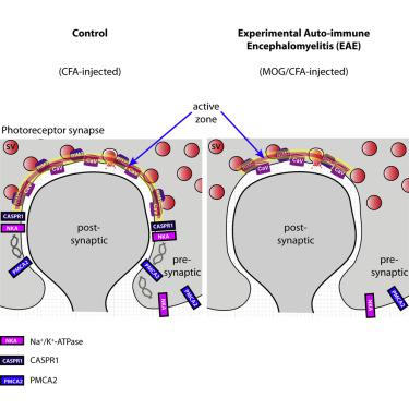
Multiple sclerosis (MS) is a demyelinating disease caused by an auto-reactive immune system. Recent studies also demonstrated synapse dysfunctions in MS patients and MS mouse models. We previously observed decreased synaptic vesicle exocytosis in photoreceptor synapses in the EAE mouse model of MS at an early, preclinical stage. In the present study, we analyzed whether synaptic defects are associated with altered presynaptic Ca2+ signaling. Using high-resolution immunolabeling, we found a reduced signal intensity of Cav-channels and RIM2 at active zones in early, preclinical EAE. In line with these morphological alterations, depolarization-evoked increases of presynaptic Ca2+ were significantly smaller. In contrast, basal presynaptic Ca2+ was elevated. We observed a decreased expression of Na+/K+-ATPase and plasma membrane Ca2+ ATPase 2 (PMCA2), but not PMCA1, in photoreceptor terminals of EAE mice that could contribute to elevated basal Ca2+. Thus, complex Ca2+ signaling alterations contribute to synaptic dysfunctions in photoreceptors in early EAE.
中文翻译:
在多发性硬化症的EAE小鼠模型中的感光细胞中突触前突触Ca 2 +信号。
多发性硬化症(MS)是由自身反应性免疫系统引起的脱髓鞘疾病。最近的研究还证明了MS患者和MS小鼠模型中的突触功能障碍。我们先前观察到,在早期的临床前阶段,MS的EAE小鼠模型中感光受体突触中的突触小泡胞吐减少。在本研究中,我们分析了突触缺陷是否与改变的突触前Ca 2+信号传导有关。使用高分辨率的免疫标记,我们发现早期临床前EAE活动区域的Cav通道和RIM2信号强度降低。与这些形态变化相一致,去极化引起的突触前Ca 2+的增加明显较小。相反,基底突触前Ca 2+被提升。我们观察到在EAE小鼠的光感受器末端,Na + / K + -ATPase和质膜Ca 2+ ATPase 2(PMCA2)的表达降低,而PMCA1没有降低,这可能导致基础Ca 2+升高。因此,复杂的Ca 2+信号转导导致早期EAE中感光器的突触功能障碍。


 京公网安备 11010802027423号