Biochimica et Biophysica Acta (BBA) - Molecular Cell Research ( IF 4.6 ) Pub Date : 2020-11-21 , DOI: 10.1016/j.bbamcr.2020.118911 Ewelina Bik , Lukasz Mateuszuk , Marta Stojak , Stefan Chlopicki , Malgorzata Baranska , Katarzyna Majzner
|
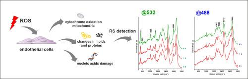
In this work, the effect of an early oxidative stress on human endothelial cells induced by menadione was studied using a combined methodology of label-free Raman imaging and fluorescence staining. Menadione-induced ROS-dependent endothelial inflammation in human aorta endothelial cells (HAEC) was studied with focus on changes in cytochrome, proteins, nucleic acids and lipids content and their distribution in cells. Fluorescence staining (ICAM-1, VCAM-1, vWF, LipidTox, MitoRos and DCF) was used to confirm endothelial inflammation and ROS generation. The results showed that short time, exposure to menadione did not cause their apoptosis or necrosis (Annexin V Apoptosis Detection Kit) within the 3 h timescale of measurement. On the other hand, 3 h of incubation, did result in endothelial inflammation (ICAM-1, VCAM-1, vWF) that was associated with an increased ROS formation (MitoRos and DCF) suggesting the oxidative stress-mediated inflammation. Chemometric analysis of spectral data enabled the determination of spectroscopic markers of menadione-induced oxidative stress–mediated endothelial inflammation including a decrease of the bands intensity of cytochrome (604, 750, 1128, 1315 and 1585 cm−1), nucleic acids bands (785 cm−1), proteins (1005 cm−1) and increased intensity of lipid bands (722, 1085, 1265, 1303, 1445 and 1660 cm−1), without changes in the spectroscopic signature of the cell nucleus. In conclusion, oxidative stress resulting in endothelial inflammation was featured by significant alterations in the number of biochemical changes in mitochondria and other cellular compartments detected by Raman spectroscopy. Most of these, coexisted with results from fluorescence imaging, and most importantly occurred earlier than the detection of increased ROS or markers of endothelial inflammation.
中文翻译:
拉曼光谱法检测甲萘醌引起的内皮炎症
在这项工作中,使用无标记拉曼成像和荧光染色的组合方法研究了早期氧化应激对甲萘醌诱导的人内皮细胞的影响。研究了甲萘醌诱导的人主动脉内皮细胞(HAEC)中的ROS依赖性内皮炎症,重点是细胞色素,蛋白质,核酸和脂质含量的变化及其在细胞中的分布。荧光染色(ICAM-1,VCAM-1,vWF,LipidTox,MitoRos和DCF)用于确认内皮炎症和ROS的产生。结果显示,短时间内,暴露于甲萘醌不会在3小时的测量范围内引起细胞凋亡或坏死(Annexin V细胞凋亡检测试剂盒)。另一方面,孵育3小时确实会导致内皮炎症(ICAM-1,VCAM-1,vWF)与ROS形成增加有关(MitoRos和DCF),表明氧化应激介导的炎症。光谱数据的化学计量分析可以测定甲萘醌诱导的氧化应激介导的内皮炎症的光谱标记,包括细胞色素(604、750、1128、1315和1585 cm-1),核酸带(785 cm -1),蛋白质(1005 cm -1)和增加的脂质带强度(722、1085、1265、1303、1445和1660 cm -1),而光谱特征没有变化细胞核。总之,导致血管内皮炎症的氧化应激的特征是线粒体和其他细胞区室的生化变化数量的显着变化(通过拉曼光谱法检测)。其中大多数与荧光成像结果共存,最重要的是比检测到升高的ROS或内皮炎症标记物更早发生。


 京公网安备 11010802027423号