当前位置:
X-MOL 学术
›
J. Biophotonics
›
论文详情
Our official English website, www.x-mol.net, welcomes your
feedback! (Note: you will need to create a separate account there.)
Hyperspectral imaging as a diagnostic tool to differentiate between amalgam tattoos and other dark pigmented intraoral lesions
Journal of Biophotonics ( IF 2.0 ) Pub Date : 2020-11-18 , DOI: 10.1002/jbio.202000424 Johannes Laimer 1 , Emanuel Bruckmoser 2 , Tom Helten 1 , Barbara Kofler 3 , Bettina Zelger 4 , Andrea Brunner 4 , Bernhard Zelger 5 , Christian W Huck 6 , Michelle Tappert 7 , Derek Rogge 7 , Michael Schirmer 8 , Johannes D Pallua 4, 9
Journal of Biophotonics ( IF 2.0 ) Pub Date : 2020-11-18 , DOI: 10.1002/jbio.202000424 Johannes Laimer 1 , Emanuel Bruckmoser 2 , Tom Helten 1 , Barbara Kofler 3 , Bettina Zelger 4 , Andrea Brunner 4 , Bernhard Zelger 5 , Christian W Huck 6 , Michelle Tappert 7 , Derek Rogge 7 , Michael Schirmer 8 , Johannes D Pallua 4, 9
Affiliation
|
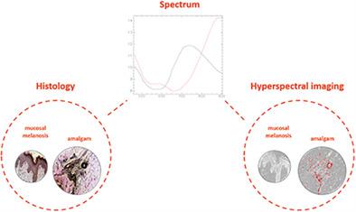
The goal of this project is to identify any in‐depth benefits and drawbacks in the diagnosis of amalgam tattoos and other pigmented intraoral lesions using hyperspectral imagery collected from amalgam tattoos, benign, and malignant melanocytic neoplasms. Software solutions capable of classifying pigmented lesions of the skin already exist, but conventional red, green and blue images may be reaching an upper limit in their performance. Emerging technologies, such as hyperspectral imaging (HSI) utilize more than a hundred, continuous data channels, while also collecting data in the infrared. A total of 18 paraffin‐embedded human tissue specimens of dark pigmented intraoral lesions (including the lip) were analyzed using visible and near‐infrared (VIS–NIR) hyperspectral imagery obtained from HE‐stained histopathological slides. Transmittance data were collected between 450 and 900 nm using a snapshot camera mounted to a microscope with a halogen light source. VIS–NIR spectra collected from different specimens, such as melanocytic cells and other tissues (eg, epithelium), produced distinct and diagnostic spectra that were used to identify these materials in several regions of interest, making it possible to distinguish between intraoral amalgam tattoos (intramucosal metallic foreign bodies) and melanocytic lesions of the intraoral mucosa and the lip (each with P < .01 using the independent t test). HSI is presented as a diagnostic tool for the rapidly growing field of digital pathology. In this preliminary study, amalgam tattoos were reliably differentiated from melanocytic lesions of the oral cavity and the lip.
中文翻译:

高光谱成像作为区分汞合金纹身和其他深色色素口腔内病变的诊断工具
该项目的目的是利用从汞齐纹身,良性和恶性黑素细胞性肿瘤中收集的高光谱图像,确定在诊断汞齐纹身和其他色素性口腔内病变方面的任何深入的利弊。能够对皮肤色素性病变进行分类的软件解决方案已经存在,但是传统的红色,绿色和蓝色图像可能已达到其性能的上限。高光谱成像(HSI)等新兴技术利用了一百多个连续的数据通道,同时还收集了红外数据。使用从HE染色的组织病理切片中获得的可见和近红外(VIS-NIR)高光谱图像,共分析了18个暗色口腔内病变(包括嘴唇)的石蜡包埋的人体组织标本。使用安装在带有卤素光源的显微镜上的快照相机在450至900 nm之间收集透射率数据。从不同样本(例如黑素细胞和其他组织(例如上皮))收集的VIS–NIR光谱产生了独特的诊断光谱,这些光谱用于在几个感兴趣的区域识别这些材料,从而可以区分口腔内的汞齐纹身(口腔内黏膜和嘴唇的黑素细胞病变和黏膜内金属异物(每个都有P <.01(使用独立t检验)。HSI被介绍为快速发展的数字病理学领域的诊断工具。在这项初步研究中,可以将汞齐纹身与口腔和嘴唇的黑素细胞病变可靠地区分开。
更新日期:2020-11-18
中文翻译:
高光谱成像作为区分汞合金纹身和其他深色色素口腔内病变的诊断工具
该项目的目的是利用从汞齐纹身,良性和恶性黑素细胞性肿瘤中收集的高光谱图像,确定在诊断汞齐纹身和其他色素性口腔内病变方面的任何深入的利弊。能够对皮肤色素性病变进行分类的软件解决方案已经存在,但是传统的红色,绿色和蓝色图像可能已达到其性能的上限。高光谱成像(HSI)等新兴技术利用了一百多个连续的数据通道,同时还收集了红外数据。使用从HE染色的组织病理切片中获得的可见和近红外(VIS-NIR)高光谱图像,共分析了18个暗色口腔内病变(包括嘴唇)的石蜡包埋的人体组织标本。使用安装在带有卤素光源的显微镜上的快照相机在450至900 nm之间收集透射率数据。从不同样本(例如黑素细胞和其他组织(例如上皮))收集的VIS–NIR光谱产生了独特的诊断光谱,这些光谱用于在几个感兴趣的区域识别这些材料,从而可以区分口腔内的汞齐纹身(口腔内黏膜和嘴唇的黑素细胞病变和黏膜内金属异物(每个都有P <.01(使用独立t检验)。HSI被介绍为快速发展的数字病理学领域的诊断工具。在这项初步研究中,可以将汞齐纹身与口腔和嘴唇的黑素细胞病变可靠地区分开。


 京公网安备 11010802027423号