当前位置:
X-MOL 学术
›
Mol. Omics
›
论文详情
Our official English website, www.x-mol.net, welcomes your
feedback! (Note: you will need to create a separate account there.)
Quantitative proteomic analysis of MDCK cell adhesion
Molecular Omics ( IF 3.0 ) Pub Date : 2020-11-12 , DOI: 10.1039/d0mo00055h Xuanqing Ye 1 , Jiamin Wang , Zilin Qiao , Di Yang , Jiao Wang , Ayimuguli Abudureyimu , Kun Yang , Yuping Feng , Zhongren Ma , Zhenbin Liu
Molecular Omics ( IF 3.0 ) Pub Date : 2020-11-12 , DOI: 10.1039/d0mo00055h Xuanqing Ye 1 , Jiamin Wang , Zilin Qiao , Di Yang , Jiao Wang , Ayimuguli Abudureyimu , Kun Yang , Yuping Feng , Zhongren Ma , Zhenbin Liu
Affiliation
|
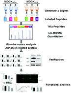
MDCK cells are a key reagent in modern vaccine production. As MDCK cells are normally adherent, creation of suspension cells for vaccine production using genetic engineering approaches is highly desirable. However, little is known regarding the mechanisms and effectors underlying MDCK cell adhesion. In this study, we performed a comparative analysis of whole protein levels between MDCK adhesion and suspension cells using an iTRAQ-based (isobaric tags for relative and absolute quantitation) proteomics approach. We found that expression of several proteins involved in cell adhesion exhibit reduced expression in suspension cells, including at the mRNA level. Proteins whose expression was reduced in suspension cells include cadherin 1 (CDH1), catenin beta-1 (CTNNB1), and catenin alpha-1 (CTNNA1), which are involved in intercellular adhesion; junction plakoglobin (JUP), desmoplakin (DSP), and desmoglein 3 (DSG3), which are desmosome components; and transglutaminase 2 (TGM2) and alpha-actinin-1 (ACTN1), which regulate the adhesion between cells and the extracellular matrix. A functional verification experiment showed that inhibition of E-cadherin significantly reduced intercellular adhesion of MDCK cells. E-Cadherin did not significantly affect the proliferation of MDCK cells and the replication of influenza virus. These findings reveal possible mechanisms underlying adhesion of MDCK cells and will guide the creation of MDCK suspension cells by genetic engineering.
中文翻译:

MDCK细胞粘附的定量蛋白质组学分析
MDCK 细胞是现代疫苗生产的关键试剂。由于 MDCK 细胞通常是贴壁细胞,因此非常需要使用基因工程方法创建用于疫苗生产的悬浮细胞。然而,关于 MDCK 细胞粘附的机制和效应器知之甚少。在这项研究中,我们使用基于 iTRAQ(用于相对和绝对定量的等压标签)蛋白质组学方法对 MDCK 粘附细胞和悬浮细胞之间的全蛋白水平进行了比较分析。我们发现参与细胞粘附的几种蛋白质的表达在悬浮细胞中表现出降低的表达,包括在 mRNA 水平。在悬浮细胞中表达降低的蛋白质包括钙粘蛋白 1 (CDH1)、连环蛋白β-1 (CTNNB1) 和连环蛋白 α-1 (CTNNA1),它们参与细胞间粘附;交界斑珠蛋白 (JUP)、桥粒蛋白 (DSP) 和桥粒芯蛋白 3 (DSG3),它们是桥粒成分;和转谷氨酰胺酶 2 (TGM2) 和 α-辅肌动蛋白-1 (ACTN1),它们调节细胞与细胞外基质之间的粘附。功能验证实验表明,抑制E-cadherin可显着降低MDCK细胞的细胞间粘附。E-Cadherin对MDCK细胞的增殖和流感病毒的复制没有显着影响。这些发现揭示了 MDCK 细胞粘附的可能机制,并将指导通过基因工程创建 MDCK 悬浮细胞。功能验证实验表明,抑制E-cadherin可显着降低MDCK细胞的细胞间粘附。E-Cadherin对MDCK细胞的增殖和流感病毒的复制没有显着影响。这些发现揭示了 MDCK 细胞粘附的可能机制,并将指导通过基因工程创建 MDCK 悬浮细胞。功能验证实验表明,抑制E-cadherin可显着降低MDCK细胞的细胞间粘附。E-Cadherin对MDCK细胞的增殖和流感病毒的复制没有显着影响。这些发现揭示了 MDCK 细胞粘附的可能机制,并将指导通过基因工程创建 MDCK 悬浮细胞。
更新日期:2020-12-09
中文翻译:
MDCK细胞粘附的定量蛋白质组学分析
MDCK 细胞是现代疫苗生产的关键试剂。由于 MDCK 细胞通常是贴壁细胞,因此非常需要使用基因工程方法创建用于疫苗生产的悬浮细胞。然而,关于 MDCK 细胞粘附的机制和效应器知之甚少。在这项研究中,我们使用基于 iTRAQ(用于相对和绝对定量的等压标签)蛋白质组学方法对 MDCK 粘附细胞和悬浮细胞之间的全蛋白水平进行了比较分析。我们发现参与细胞粘附的几种蛋白质的表达在悬浮细胞中表现出降低的表达,包括在 mRNA 水平。在悬浮细胞中表达降低的蛋白质包括钙粘蛋白 1 (CDH1)、连环蛋白β-1 (CTNNB1) 和连环蛋白 α-1 (CTNNA1),它们参与细胞间粘附;交界斑珠蛋白 (JUP)、桥粒蛋白 (DSP) 和桥粒芯蛋白 3 (DSG3),它们是桥粒成分;和转谷氨酰胺酶 2 (TGM2) 和 α-辅肌动蛋白-1 (ACTN1),它们调节细胞与细胞外基质之间的粘附。功能验证实验表明,抑制E-cadherin可显着降低MDCK细胞的细胞间粘附。E-Cadherin对MDCK细胞的增殖和流感病毒的复制没有显着影响。这些发现揭示了 MDCK 细胞粘附的可能机制,并将指导通过基因工程创建 MDCK 悬浮细胞。功能验证实验表明,抑制E-cadherin可显着降低MDCK细胞的细胞间粘附。E-Cadherin对MDCK细胞的增殖和流感病毒的复制没有显着影响。这些发现揭示了 MDCK 细胞粘附的可能机制,并将指导通过基因工程创建 MDCK 悬浮细胞。功能验证实验表明,抑制E-cadherin可显着降低MDCK细胞的细胞间粘附。E-Cadherin对MDCK细胞的增殖和流感病毒的复制没有显着影响。这些发现揭示了 MDCK 细胞粘附的可能机制,并将指导通过基因工程创建 MDCK 悬浮细胞。


 京公网安备 11010802027423号