当前位置:
X-MOL 学术
›
Microsc. Res. Tech.
›
论文详情
Our official English website, www.x-mol.net, welcomes your
feedback! (Note: you will need to create a separate account there.)
Morphology of the male and female reproductive tracts of virgin and mated Chrysoperla externa (Hagen, 1861) (Neuroptera: Chrysopidae)
Microscopy Research and Technique ( IF 2.0 ) Pub Date : 2020-11-15 , DOI: 10.1002/jemt.23645 Priscylla Costa Dantas 1 , Helen Cristina Pinto Santos 2 , Hakan Bozdoğan 3 , José Eduardo Serrão 4 , Geraldo Andrade Carvalho 1
Microscopy Research and Technique ( IF 2.0 ) Pub Date : 2020-11-15 , DOI: 10.1002/jemt.23645 Priscylla Costa Dantas 1 , Helen Cristina Pinto Santos 2 , Hakan Bozdoğan 3 , José Eduardo Serrão 4 , Geraldo Andrade Carvalho 1
Affiliation
|
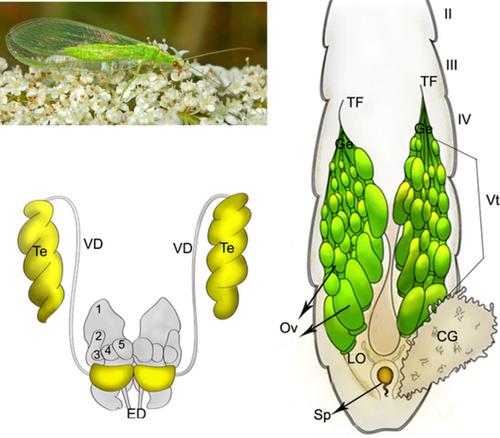
Predatory insects have reproductive organs rich in complex changes that may be responsible for the success of their population growth. The species Chrysoperla externa is a predator used in biological control programs in Latin America. However, there is no morphological data about the morphology of the reproductive tract in this insect. This study describes the morphology of the reproductive organs of virgin and mated C. externa male and female. The male has a pair of testes yellow in color and five pairs of accessory glands closely associated with the seminal vesicles. The testis follicles are twisted filled with cysts in different developmental stages. The pair of ovaries in the females shows asymmetry with 9–11 ovarioles per ovary with oocytes in different developmental stages and a spherical spermatheca. Virgin and mated males have no differences in the size of the testes, seminal vesicle, and accessory glands. C. externa females show morphological changes in the reproductive tract according to sexual maturation, which is triggered by mating. The ovary activation occurs after female mating. The ovaries are of merotistic polytrophic type. The spermathecal reservoir is lined by a flattened epithelium with a thin cuticular intima and associated with well‐developed muscles. It is concluded that the reproductive tract of C. externa is similar in virgin and mated males and females. Egg production is activated only after mating and the development of reproductive tract structures is faster in mated females.
中文翻译:

原始和已交配的外层金藻雄性和雌性生殖道的形态学 (Hagen, 1861) (Neuroptera: Chrysopidae)
捕食性昆虫的生殖器官具有丰富的复杂变化,这可能是其种群增长成功的原因。Chrysoperla externa物种是拉丁美洲生物控制计划中使用的捕食者。然而,没有关于这种昆虫生殖道形态的形态学数据。这项研究描述了未婚和已交配的C生殖器官的形态。外部男性和女性。雄性有一对黄色的睾丸和五对与精囊密切相关的附属腺体。在不同的发育阶段,睾丸的卵泡是扭曲的,充满了囊肿。雌性的一对卵巢表现出不对称性,每个卵巢有 9-11 个卵巢,卵母细胞处于不同发育阶段,并且有一个球形的受精囊。处女和已交配的雄性在睾丸、精囊和附属腺体的大小方面没有差异。Ç。外部根据交配引发的性成熟,雌性在生殖道中表现出形态变化。卵巢激活发生在雌性交配后。卵巢是分裂多养型。受精囊内衬有扁平的上皮,内膜薄,与发达的肌肉相关。得出的结论是,C的生殖道。外在处女和已交配的雄性和雌性相似。产卵只有在交配后才会被激活,并且交配后的雌性生殖道结构的发育更快。
更新日期:2020-11-15
中文翻译:
原始和已交配的外层金藻雄性和雌性生殖道的形态学 (Hagen, 1861) (Neuroptera: Chrysopidae)
捕食性昆虫的生殖器官具有丰富的复杂变化,这可能是其种群增长成功的原因。Chrysoperla externa物种是拉丁美洲生物控制计划中使用的捕食者。然而,没有关于这种昆虫生殖道形态的形态学数据。这项研究描述了未婚和已交配的C生殖器官的形态。外部男性和女性。雄性有一对黄色的睾丸和五对与精囊密切相关的附属腺体。在不同的发育阶段,睾丸的卵泡是扭曲的,充满了囊肿。雌性的一对卵巢表现出不对称性,每个卵巢有 9-11 个卵巢,卵母细胞处于不同发育阶段,并且有一个球形的受精囊。处女和已交配的雄性在睾丸、精囊和附属腺体的大小方面没有差异。Ç。外部根据交配引发的性成熟,雌性在生殖道中表现出形态变化。卵巢激活发生在雌性交配后。卵巢是分裂多养型。受精囊内衬有扁平的上皮,内膜薄,与发达的肌肉相关。得出的结论是,C的生殖道。外在处女和已交配的雄性和雌性相似。产卵只有在交配后才会被激活,并且交配后的雌性生殖道结构的发育更快。


 京公网安备 11010802027423号