当前位置:
X-MOL 学术
›
Clin. Exp. Immunol.
›
论文详情
Our official English website, www.x-mol.net, welcomes your
feedback! (Note: you will need to create a separate account there.)
Roles of cytokines and T cells in the pathogenesis of myasthenia gravis
Clinical & Experimental Immunology ( IF 3.4 ) Pub Date : 2020-11-13 , DOI: 10.1111/cei.13546 A Uzawa 1 , S Kuwabara 1 , S Suzuki 2 , T Imai 3 , H Murai 4 , Y Ozawa 1 , M Yasuda 1 , Y Nagane 5 , K Utsugisawa 5
Clinical & Experimental Immunology ( IF 3.4 ) Pub Date : 2020-11-13 , DOI: 10.1111/cei.13546 A Uzawa 1 , S Kuwabara 1 , S Suzuki 2 , T Imai 3 , H Murai 4 , Y Ozawa 1 , M Yasuda 1 , Y Nagane 5 , K Utsugisawa 5
Affiliation
|
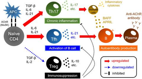
Myasthenia gravis (MG) is characterized by muscle weakness and fatigue caused by the presence of autoantibodies against the acetylcholine receptor (AChR) or the muscle‐specific tyrosine kinase (MuSK). Activated T, B and plasma cells, as well as cytokines, play important roles in the production of pathogenic autoantibodies and the induction of inflammation at the neuromuscular junction in MG. Many studies have focused on the role of cytokines and lymphocytes in anti‐AChR antibody‐positive MG. Chronic inflammation mediated by T helper type 17 (Th17) cells, the promotion of autoantibody production from B cells and plasma cells by follicular Th (Tfh) cells and the activation of the immune response by dysfunction of regulatory T (Treg) cells may contribute to the exacerbation of the MG pathogenesis. In fact, an increased number of Th17 cells and Tfh cells and dysfunction of Treg cells have been reported in patients with anti‐AChR antibody‐positive MG; moreover, the number of these cells was correlated with clinical parameters in patients with MG. Regarding cytokines, interleukin (IL)‐17; a Th17‐related cytokine, IL‐21 (a Tfh‐related cytokine), the B‐cell‐activating factor (BAFF; a B cell‐related cytokine) and a proliferation‐inducing ligand (APRIL; a B cell‐related cytokine) have been reported to be up‐regulated and associated with clinical parameters of MG. This review focuses on the current understanding of the involvement of cytokines and lymphocytes in the immunological pathogenesis of MG, which may lead to the development of novel therapies for this disease in the near future.
中文翻译:

细胞因子和T细胞在重症肌无力发病机制中的作用
重症肌无力 (MG) 的特征是由于存在针对乙酰胆碱受体 (AChR) 或肌肉特异性酪氨酸激酶 (MuSK) 的自身抗体而导致的肌肉无力和疲劳。活化的 T、B 和浆细胞以及细胞因子在 MG 中致病性自身抗体的产生和神经肌肉接头炎症的诱导中起重要作用。许多研究都集中在细胞因子和淋巴细胞在抗 AChR 抗体阳性 MG 中的作用。由 T 辅助 17 型 (Th17) 细胞介导的慢性炎症、滤泡 Th (Tfh) 细胞促进 B 细胞和浆细胞产生自身抗体以及调节性 T (T reg ) 功能障碍激活免疫反应) 细胞可能导致 MG 发病机制的恶化。事实上,Th17 细胞和 Tfh 细胞数量增加以及 T reg功能障碍在抗 AChR 抗体阳性的 MG 患者中报告了细胞;此外,这些细胞的数量与 MG 患者的临床参数相关。关于细胞因子,白细胞介素 (IL)-17;Th17 相关细胞因子、IL-21(Tfh 相关细胞因子)、B 细胞激活因子(BAFF;B 细胞相关细胞因子)和增殖诱导配体(APRIL;B 细胞相关细胞因子)已报道上调并与 MG 的临床参数相关。本综述着重于目前对细胞因子和淋巴细胞参与 MG 免疫发病机制的了解,这可能会导致在不久的将来开发针对该疾病的新疗法。
更新日期:2020-11-13
中文翻译:
细胞因子和T细胞在重症肌无力发病机制中的作用
重症肌无力 (MG) 的特征是由于存在针对乙酰胆碱受体 (AChR) 或肌肉特异性酪氨酸激酶 (MuSK) 的自身抗体而导致的肌肉无力和疲劳。活化的 T、B 和浆细胞以及细胞因子在 MG 中致病性自身抗体的产生和神经肌肉接头炎症的诱导中起重要作用。许多研究都集中在细胞因子和淋巴细胞在抗 AChR 抗体阳性 MG 中的作用。由 T 辅助 17 型 (Th17) 细胞介导的慢性炎症、滤泡 Th (Tfh) 细胞促进 B 细胞和浆细胞产生自身抗体以及调节性 T (T reg ) 功能障碍激活免疫反应) 细胞可能导致 MG 发病机制的恶化。事实上,Th17 细胞和 Tfh 细胞数量增加以及 T reg功能障碍在抗 AChR 抗体阳性的 MG 患者中报告了细胞;此外,这些细胞的数量与 MG 患者的临床参数相关。关于细胞因子,白细胞介素 (IL)-17;Th17 相关细胞因子、IL-21(Tfh 相关细胞因子)、B 细胞激活因子(BAFF;B 细胞相关细胞因子)和增殖诱导配体(APRIL;B 细胞相关细胞因子)已报道上调并与 MG 的临床参数相关。本综述着重于目前对细胞因子和淋巴细胞参与 MG 免疫发病机制的了解,这可能会导致在不久的将来开发针对该疾病的新疗法。


 京公网安备 11010802027423号