当前位置:
X-MOL 学术
›
Biomater. Sci.
›
论文详情
Our official English website, www.x-mol.net, welcomes your feedback! (Note: you will need to create a separate account there.)
In vivo evaluation of a pro-healing polydopamine coated stent through an in-stent restenosis rat model
Biomaterials Science ( IF 6.6 ) Pub Date : 2020-10-22 , DOI: 10.1039/d0bm01204a Adrien Hertault 1, 2, 3, 4, 5 , Feng Chai 1, 2, 3, 4, 5 , Mickael Maton 1, 2, 3, 4, 5 , Jonathan Sobocinski 1, 2, 3, 4, 5 , Patrice Woisel 1, 6, 7, 8, 9 , Blandine Maurel 2, 10, 11, 12, 13 , Joël Lyskawa 1, 6, 7, 8, 9 , Nicolas Blanchemain 1, 2, 3, 4, 5
Biomaterials Science ( IF 6.6 ) Pub Date : 2020-10-22 , DOI: 10.1039/d0bm01204a Adrien Hertault 1, 2, 3, 4, 5 , Feng Chai 1, 2, 3, 4, 5 , Mickael Maton 1, 2, 3, 4, 5 , Jonathan Sobocinski 1, 2, 3, 4, 5 , Patrice Woisel 1, 6, 7, 8, 9 , Blandine Maurel 2, 10, 11, 12, 13 , Joël Lyskawa 1, 6, 7, 8, 9 , Nicolas Blanchemain 1, 2, 3, 4, 5
Affiliation
|
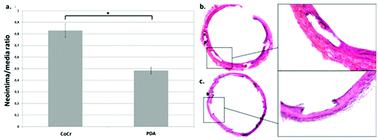
Drug-eluting stents have demonstrated efficiency in in-stent restenosis (ISR) but induced a risk of late acute thrombosis by delaying strut re-endothelialization. Polydopamine (PDA), a biocompatible polymer inspired from adhesive proteins of mussels, has been reported to promote endothelial cell (EC) proliferation while limiting SMC proliferation in vitro, thus suggesting the pro-healing potential. This study aimed at evaluating in vivo the impact of the pro-healing PDA-coated stent on ISR and on the quality of the strut re-endothelialization in a rat model. PDA-coated stents demonstrated a significant reduction in ISR in vivo compared to bare metal stents (ratio neointima/media = 0.48 (±0.26) versus 0.83 (±0.42), p < 0.001). Western blot analyses identified a trend towards an increased activation of p38 MAPK phosphorylation and its anti-proliferative effects on vascular SMC that could explain the results observed in morphological analyses. This bioinspired and biocompatible polydopamine layer could intrinsically limit ISR. In addition, according to its latent reactivity, PDA offers the possibility to immobilize some relevant drugs on the PDA-functionalized stent to provide potential synergistic effects.
中文翻译:

通过支架内再狭窄大鼠模型对愈合的聚多巴胺涂层支架进行体内评估
药物洗脱支架在支架内再狭窄(ISR)中已证明有效,但通过延迟支杆再内皮化而引起晚期急性血栓形成的风险。聚多巴胺(PDA)是一种受贻贝粘附蛋白启发的生物相容性聚合物,据报道可促进内皮细胞(EC)增殖,同时限制体外SMC增殖,因此表明其具有修复的潜力。这项研究旨在评估在大鼠模型中,愈合前PDA涂层支架对ISR的影响以及支杆再内皮化的质量。与裸金属支架相比,PDA涂层支架在体内的ISR明显降低(新内膜/介质比= 0.48(±0.26)对0.83(±0.42),p<0.001)。Western印迹分析确定了p38 MAPK磷酸化激活增加的趋势及其对血管SMC的抗增殖作用,这可以解释在形态分析中观察到的结果。这种具有生物启发性和生物相容性的聚多巴胺层可以从本质上限制ISR。此外,根据其潜在的反应性,PDA还提供了将一些相关药物固定在PDA功能化支架上的可能性,以提供潜在的协同作用。
更新日期:2020-11-12
中文翻译:
通过支架内再狭窄大鼠模型对愈合的聚多巴胺涂层支架进行体内评估
药物洗脱支架在支架内再狭窄(ISR)中已证明有效,但通过延迟支杆再内皮化而引起晚期急性血栓形成的风险。聚多巴胺(PDA)是一种受贻贝粘附蛋白启发的生物相容性聚合物,据报道可促进内皮细胞(EC)增殖,同时限制体外SMC增殖,因此表明其具有修复的潜力。这项研究旨在评估在大鼠模型中,愈合前PDA涂层支架对ISR的影响以及支杆再内皮化的质量。与裸金属支架相比,PDA涂层支架在体内的ISR明显降低(新内膜/介质比= 0.48(±0.26)对0.83(±0.42),p<0.001)。Western印迹分析确定了p38 MAPK磷酸化激活增加的趋势及其对血管SMC的抗增殖作用,这可以解释在形态分析中观察到的结果。这种具有生物启发性和生物相容性的聚多巴胺层可以从本质上限制ISR。此外,根据其潜在的反应性,PDA还提供了将一些相关药物固定在PDA功能化支架上的可能性,以提供潜在的协同作用。


 京公网安备 11010802027423号