Our official English website, www.x-mol.net, welcomes your
feedback! (Note: you will need to create a separate account there.)
Quantification of the carbon bonding state in amorphous carbon materials: a comparison between EELS and NEXAFS measurements
Carbon ( IF 10.5 ) Pub Date : 2021-03-01 , DOI: 10.1016/j.carbon.2020.11.021 Filippo Mangolini , Zixuan Li , Matthew A. Marcus , Reinhard Schneider , Martin Dienwiebel
Carbon ( IF 10.5 ) Pub Date : 2021-03-01 , DOI: 10.1016/j.carbon.2020.11.021 Filippo Mangolini , Zixuan Li , Matthew A. Marcus , Reinhard Schneider , Martin Dienwiebel
|
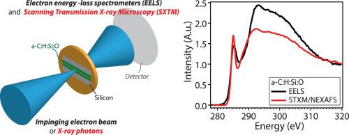
The quantitative determination of the carbon hybridization is critical for establishing processing-structure-properties relationships for carbon-based materials, including amorphous carbon coatings. While several techniques have been employed to characterize the amount of sp$^{2}$ and sp$^{3}$ carbon in these materials, direct comparisons between analytical results are limited. Here, we compare near edge X-rayabsorptionfine structure (NEXAFS) spectra of a silicon- and oxygen-containing hydrogenated amorphouscarbon (a-C:H:Si:O) coating acquired in synchrotron-based scanning transmission X-ray microscopy (STXM) mode with electron energy loss spectra (EELS) obtained from the same a-C:H:Si:O lamella. While the fractions of sp$^{2}$ carbon computed from STXM and EELS spectra are in close agreement, the comparison of NEXAFS spectra acquired in STXM mode with NEXAFS spectra collected in partial electron yield mode on a flat a-C:H:Si:O surface indicated that the destructive preparation of thin lamellae for STXM analyses induces variations in the structure of a-C:H:Si:O, namely the breakage of carbon-siliconand carbon-hydrogen bonds, a change in ordering of sp$^{2}$-bonded carbon, and an increase in the sp$^{2}$ carbon fraction. These findings can help scientists in the careful interpretation of spectroscopic results obtained from the analysis of samples made of metastable materials after the destructive preparation ofspecimens for analytical purposes.
中文翻译:

无定形碳材料中碳键状态的量化:EELS 和 NEXAFS 测量之间的比较
碳杂化的定量测定对于建立碳基材料(包括无定形碳涂层)的加工-结构-性能关系至关重要。虽然已采用多种技术来表征这些材料中 sp$^{2}$ 和 sp$^{3}$ 碳的量,但分析结果之间的直接比较是有限的。在这里,我们比较了在基于同步加速器的扫描透射 X 射线显微镜 (STXM) 模式中获得的含硅和氧的氢化非晶碳 (aC:H:Si:O) 涂层的近边缘 X 射线吸收精细结构 (NEXAFS) 光谱与从相同的 aC:H:Si:O 薄片获得的电子能量损失谱 (EELS)。虽然从 STXM 和 EELS 光谱计算的 sp$^{2}$ 碳的分数非常一致,在 STXM 模式下获得的 NEXAFS 光谱与在平坦的 aC:H:Si:O 表面上以部分电子产率模式收集的 NEXAFS 光谱的比较表明,用于 STXM 分析的薄层的破坏性制备导致了 aC:H 结构的变化: Si:O,即碳-硅和碳-氢键断裂,sp$^{2}$-键合碳的顺序发生变化,sp$^{2}$碳分数增加。这些发现可以帮助科学家仔细解释光谱结果,这些结果是在对样品进行破坏性制备以进行分析后对亚稳态材料制成的样品进行分析而获得的。O 表面表明用于 STXM 分析的薄层的破坏性制备导致 aC:H:Si:O 结构的变化,即碳 - 硅和碳 - 氢键的断裂,sp$^{2} 的顺序发生变化$-键合碳,以及 sp$^{2}$ 碳分数的增加。这些发现可以帮助科学家仔细解释光谱结果,这些结果是在对样品进行破坏性制备以进行分析后对亚稳态材料制成的样品进行分析而获得的。O 表面表明,用于 STXM 分析的薄层的破坏性制备导致 aC:H:Si:O 结构的变化,即碳 - 硅和碳 - 氢键的断裂,sp$^{2} 的顺序发生变化$-键合碳,以及 sp$^{2}$ 碳分数的增加。这些发现可以帮助科学家仔细解释光谱结果,这些结果是在对样品进行破坏性制备以进行分析后对亚稳态材料制成的样品进行分析而获得的。
更新日期:2021-03-01
中文翻译:
无定形碳材料中碳键状态的量化:EELS 和 NEXAFS 测量之间的比较
碳杂化的定量测定对于建立碳基材料(包括无定形碳涂层)的加工-结构-性能关系至关重要。虽然已采用多种技术来表征这些材料中 sp$^{2}$ 和 sp$^{3}$ 碳的量,但分析结果之间的直接比较是有限的。在这里,我们比较了在基于同步加速器的扫描透射 X 射线显微镜 (STXM) 模式中获得的含硅和氧的氢化非晶碳 (aC:H:Si:O) 涂层的近边缘 X 射线吸收精细结构 (NEXAFS) 光谱与从相同的 aC:H:Si:O 薄片获得的电子能量损失谱 (EELS)。虽然从 STXM 和 EELS 光谱计算的 sp$^{2}$ 碳的分数非常一致,在 STXM 模式下获得的 NEXAFS 光谱与在平坦的 aC:H:Si:O 表面上以部分电子产率模式收集的 NEXAFS 光谱的比较表明,用于 STXM 分析的薄层的破坏性制备导致了 aC:H 结构的变化: Si:O,即碳-硅和碳-氢键断裂,sp$^{2}$-键合碳的顺序发生变化,sp$^{2}$碳分数增加。这些发现可以帮助科学家仔细解释光谱结果,这些结果是在对样品进行破坏性制备以进行分析后对亚稳态材料制成的样品进行分析而获得的。O 表面表明用于 STXM 分析的薄层的破坏性制备导致 aC:H:Si:O 结构的变化,即碳 - 硅和碳 - 氢键的断裂,sp$^{2} 的顺序发生变化$-键合碳,以及 sp$^{2}$ 碳分数的增加。这些发现可以帮助科学家仔细解释光谱结果,这些结果是在对样品进行破坏性制备以进行分析后对亚稳态材料制成的样品进行分析而获得的。O 表面表明,用于 STXM 分析的薄层的破坏性制备导致 aC:H:Si:O 结构的变化,即碳 - 硅和碳 - 氢键的断裂,sp$^{2} 的顺序发生变化$-键合碳,以及 sp$^{2}$ 碳分数的增加。这些发现可以帮助科学家仔细解释光谱结果,这些结果是在对样品进行破坏性制备以进行分析后对亚稳态材料制成的样品进行分析而获得的。


 京公网安备 11010802027423号