Brain Research ( IF 2.9 ) Pub Date : 2020-11-07 , DOI: 10.1016/j.brainres.2020.147201 T Akgul Caglar 1 , Z B Durdu 2 , M U Turhan 3 , M Y Gunal 4 , M S Aydın 5 , G Ozturk 6 , E Cagavi 7
|
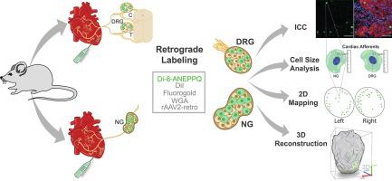
The identity of sensory neurons innervating the heart tissue and the extent of information reported to the brain via these neurons are poorly understood. In order to evaluate the multidimensional distribution and abundance of the cardiac spinal and vagal afferents, we assessed the retrograde labeling efficiency of various tracers, and mapped the cardiac afferents qualitatively and quantitatively at the bilateral nodose ganglia (NGs) and dorsal root ganglia (DRGs). From the five different retrograde tracers evaluated, Di-8-ANEPPQ yielded reproducibly the highest labeling efficiency of cardiac afferents. We demonstrated specific cardiac afferents at NGs and C4 to T11 DRG segments. Next, the 2D reconstruction of the tissue sections and 3D imaging of the whole NGs and DRGs revealed homogeneous and bilateral distribution of cardiac afferents. The quantitative analyses of the labeled cardiac afferents demonstrated approximately 5–6% of the soma in NGs that were equally distributed bilaterally. The neuronal character of Di-8-ANEPPQ labeled cells were validated by coimmunostaning with pan-neuronal marker Tuj-1. In addition, the cell diameters of labeled cardiac sensory neurons were found smaller than 20 μm, implying the nociceptor phenotype confirmed by co-labeling with TRPV1 and Di-8-ANEPPQ. Importantly, co-labeling with two distinct tracers Di-8-ANEPPQ and WGA-647 demonstrated exclusively the same cardiac afferents in DRGs and NGs, validating our findings. Collectively, our findings revealed the cardiac afferents in NGs bilaterally and DRGs with the highest labeling efficiency reported, spatial distribution and quantitation at both 2D and 3D levels, furthering our understanding of this novel neuron population.
中文翻译:
通过逆行标记评估脊髓和迷走神经节双侧心脏传入分布
支配心脏组织的感觉神经元的身份以及通过这些神经元向大脑报告的信息范围知之甚少。为了评估心脏脊髓和迷走神经传入神经的多维分布和丰度,我们评估了各种示踪剂的逆行标记效率,并在双侧结节神经节(NGs)和背根神经节(DRGs)处定性和定量地绘制了心脏传入神经。 . 从评估的五种不同的逆行示踪剂中,Di-8-ANEPPQ 可重复产生最高的心脏传入标记效率。我们在 NGs 和 C4 到 T11 DRG 段展示了特定的心脏传入。接下来,组织切片的 2D 重建和整个 NG 和 DRG 的 3D 成像揭示了心脏传入的均匀和双侧分布。标记的心脏传入的定量分析表明,NGs 中大约 5-6% 的体细胞在双侧均匀分布。Di-8-ANEPPQ 标记细胞的神经元特征通过与泛神经元标记 Tuj-1 的共免疫染色来验证。此外,发现标记的心脏感觉神经元的细胞直径小于 20 μm,这意味着通过与 TRPV1 和 Di-8-ANEPPQ 共标记证实了伤害感受器表型。重要的是,用两种不同的示踪剂 Di-8-ANEPPQ 和 WGA-647 共同标记仅在 DRG 和 NG 中证明了相同的心脏传入,验证了我们的发现。总的来说,我们的研究结果揭示了双侧 NG 和 DRG 中的心脏传入,报告的标记效率最高,空间分布和 2D 和 3D 水平的定量,


 京公网安备 11010802027423号