Acta Biomaterialia ( IF 9.4 ) Pub Date : 2020-11-07 , DOI: 10.1016/j.actbio.2020.11.006 Yu Jung Shin , Ryan T. Shafranek , Jonathan H. Tsui , Jelisha Walcott , Alshakim Nelson , Deok-Ho Kim
|
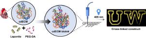
3D bioprinting is a powerful technique for engineering tissues used to study cell behavior and tissue properties in vitro. With the right formulation and printing parameters, bioinks can provide native biological and mechanical cues while allowing for versatile 3D structures that recapitulate tissue-level organization. Bio-based materials that support cellular adhesion, differentiation, and proliferation - including gelatin, collagen, hyaluronic acid, and alginate - have been successfully used as bioinks. In particular, decellularized extracellular matrix (dECM) has become a promising material with the unique ability to maintain both biochemical and topographical micro-environments of native tissues. However, dECM has shown technical limitations for 3D printing (3DP) applications posed by its intrinsically low mechanical stability. Herein, we report hydrogel bioinks composed of partially digested, porcine cardiac decellularized extracellular matrix (cdECM), Laponite-XLG nanoclay, and poly(ethylene glycol)-diacrylate (PEG-DA). The Laponite facilitated extrusion-based 3DP, while PEG-DA enabled photo-polymerization after printing. Improving upon previously reported bioinks derived from dECM, our bioinks combine extrudability, shape fidelity, rapid cross-linking, and cytocompatibility in a single formulation (> 97% viability of encapsulated human cardiac fibroblasts and > 94% viability of human induced pluripotent stem cell derived cardiomyocytes after 7 days). The compressive modulus of the cured hydrogel bioinks was tunable from 13.4-89 kPa by changing the concentration of PEG-DA in the bioink formulation. Importantly, this span of mechanical stiffness encompasses ranges of tissue stiffness from healthy (compressive modulus ~5-15 kPa) to fibrotic (compressive modulus ~30-100 kPa) cardiac tissue states. The printed constructs demonstrated shape fidelity, adaptability to different printing conditions, and high cell viability following extrusion and photo-polymerization, highlighting the potential for applications in modeling both healthy and fibrotic cardiac tissue.
中文翻译:
从心脏脱细胞的细胞外基质衍生出来的机械调谐生物墨水的3D生物打印
3D生物打印是一种工程化组织的强大技术,可用于体外研究细胞行为和组织特性。借助正确的配方和印刷参数,生物墨水可以提供天然的生物学和机械线索,同时允许通用的3D结构来概括组织级组织。支持细胞粘附,分化和增殖的基于生物的材料(包括明胶,胶原蛋白,透明质酸和藻酸盐)已成功用作生物墨水。特别是,脱细胞的细胞外基质(dECM)已成为一种有前途的材料,具有维持天然组织生化和地形学微环境的独特能力。但是,dECM因其固有的低机械稳定性而表现出对3D打印(3DP)应用的技术限制。在此,我们报道了由部分消化的猪心脏脱细胞细胞外基质(cdECM)组成的水凝胶生物墨水,Laponite-XLG纳米粘土和聚(乙二醇)-二丙烯酸酯(PEG-DA)。Laponite促进了基于挤出的3DP,而PEG-DA可以在印刷后进行光聚合。在以前报道的源自dECM的生物墨水的基础上进行改进,我们的生物墨水在单一配方中结合了可挤出性,形状保真度,快速交联和细胞相容性(> 97%的封装人心脏成纤维细胞活力和> 94%的人诱导多能干细胞活力) 7天后的心肌细胞)。通过改变生物墨水配方中PEG-DA的浓度,可将固化的水凝胶生物墨水的压缩模量从13.4-89 kPa可调。重要的,机械刚度的范围涵盖了从健康(压缩模量约5-15 kPa)到纤维化(压缩模量约30-100 kPa)心脏组织状态的组织刚度范围。印刷的构造物表现出形状保真度,对不同印刷条件的适应性以及挤出和光聚合后的高细胞活力,突出了在健康和纤维化心脏组织建模中的应用潜力。


 京公网安备 11010802027423号