Journal of Neuroimmune Pharmacology ( IF 5.2 ) Pub Date : 2020-11-09 , DOI: 10.1007/s11481-020-09969-w Jiaofei Zhang 1, 2 , Hui Li 1 , Hao Yang 3 , Jianhua Lin 4 , You Wang 4 , Qianjun Zhang 2 , Wei-Qiang Gao 1, 5 , Huiming Xu 1
|
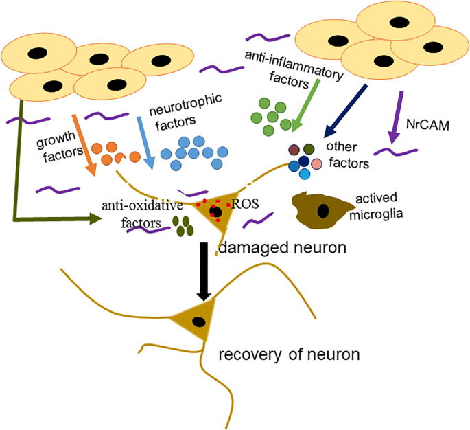
Human amniotic epithelial cells (hAECs) have been reported to have neuroprotective roles in Parkinson’s disease (PD) animal models. However, the molecular mechanism is not fully understood. The present study was designed to explore the possible mechanism by which hAECs ameliorate PD symptoms and the important paracrine factors produced by hAECs that attribute to the recovery of dopaminergic neurons. Thus, we performed in vivo and in vitro experiments with hAECs in PD models or lesioned dopaminergic neurons, respectively. First, hAECs were transplanted into the striatum of 1-methyl-4-phenyl-1,2,3,6-tetrahydropyridine (MPTP)-induced PD mice and motor deficits were significantly attenuated. Second, the grafts prevented the loss of nigral dopaminergic neurons and promoted the outgrowth of neurites and striatal axon fibers in PD mice. In addition, decreased microglial activation, inflammatory factor levels and MPTP-induced excessive reactive oxygen species (ROS) levels were also observed in hAEC-treated PD mice. In vitro, we found that the conditioned medium (CM) from hAECs promoted the survival of mesencephalic dopaminergic neurons stimulated with 1-methyl-4-phenylpyridine (MPP+) and induced neurite outgrowth. Next, analysis of hAEC-CM with an antibody array of 507 soluble target proteins revealed that the levels of many neurotrophic factors, growth factors, neuronal cell adhesion molecule (NrCAM) and anti-inflammatory factors were evidently high. In addition, antibody neutralization experiments showed that many of these factors contributed to the survival and growth of dopaminergic neurons and neurite outgrowth. More importantly, we found that the anti-inflammatory factor interleukin-1 receptor antagonist (IL-1ra) also augmented the survival of dopaminergic neurons, demonstrating for the first time an anti-oxidative and anti-inflammatory role of hAECs in PD mice, which represents a novel molecular mechanism of hAECs in the treatment of PD.
中文翻译:
人羊膜上皮细胞主要通过神经保护、抗氧化和抗炎因子缓解小鼠帕金森病模型
据报道,人类羊膜上皮细胞 (hAEC) 在帕金森病 (PD) 动物模型中具有神经保护作用。然而,分子机制尚不完全清楚。本研究旨在探讨 hAECs 改善 PD 症状的可能机制以及 hAECs 产生的重要旁分泌因子,这些因子归因于多巴胺能神经元的恢复。因此,我们分别在 PD 模型或受损的多巴胺能神经元中对 hAEC 进行了体内和体外实验。首先,将 hAEC 移植到 1-甲基-4-苯基-1,2,3,6-四氢吡啶 (MPTP) 诱导的 PD 小鼠的纹状体中,运动缺陷显着减弱。其次,移植物防止了 PD 小鼠黑质多巴胺能神经元的损失,并促进了神经突和纹状体轴突纤维的生长。此外,在 hAEC 治疗的 PD 小鼠中也观察到小胶质细胞活化、炎症因子水平和 MPTP 诱导的过量活性氧 (ROS) 水平降低。体外,我们发现来自 hAECs 的条件培养基 (CM) 促进了用 1-甲基-4-苯基吡啶 (MPP +) 并诱导神经突生长。接下来,用 507 种可溶性靶蛋白的抗体阵列分析 hAEC-CM,发现许多神经营养因子、生长因子、神经元细胞粘附分子 (NrCAM) 和抗炎因子的水平明显较高。此外,抗体中和实验表明,许多这些因素有助于多巴胺能神经元的存活和生长以及神经突的生长。更重要的是,我们发现抗炎因子白细胞介素 1 受体拮抗剂 (IL-1ra) 也增加了多巴胺能神经元的存活率,首次证明了 hAEC 在 PD 小鼠中的抗氧化和抗炎作用。代表了hAECs治疗PD的新分子机制。


 京公网安备 11010802027423号