当前位置:
X-MOL 学术
›
Dev. Growth Differ.
›
论文详情
Our official English website, www.x-mol.net, welcomes your
feedback! (Note: you will need to create a separate account there.)
Light‐sheet microscopy‐based 3D single‐cell tracking reveals a correlation between cell cycle and the start of endoderm cell internalization in early zebrafish development
Development, Growth & Differentiation ( IF 1.7 ) Pub Date : 2020-11-03 , DOI: 10.1111/dgd.12695 Akiko Kondow 1, 2 , Kiyoshi Ohnuma 3, 4 , Yasuhiro Kamei 5, 6 , Atsushi Taniguchi 2, 7 , Ryoma Bise 8 , Yoichi Sato 9 , Hisateru Yamaguchi 1 , Shigenori Nonaka 2, 5, 6 , Keiichiro Hashimoto 1
Development, Growth & Differentiation ( IF 1.7 ) Pub Date : 2020-11-03 , DOI: 10.1111/dgd.12695 Akiko Kondow 1, 2 , Kiyoshi Ohnuma 3, 4 , Yasuhiro Kamei 5, 6 , Atsushi Taniguchi 2, 7 , Ryoma Bise 8 , Yoichi Sato 9 , Hisateru Yamaguchi 1 , Shigenori Nonaka 2, 5, 6 , Keiichiro Hashimoto 1
Affiliation
|
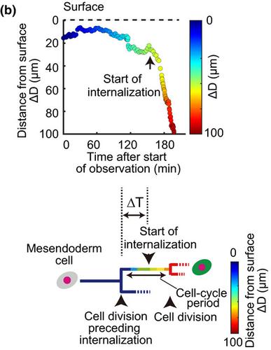
Controlling the initiation of cell migration plays a fundamental role in shaping the tissue during embryonic development. During gastrulation in zebrafish, some mesendoderm cells migrate inward to form the endoderm as the innermost germ layer along the yolk syncytial layer. However, how the initiation of inward migration is regulated is poorly understood. In this study, we performed light‐sheet microscopy‐based 3D single‐cell tracking consisting of (a) whole‐embryo time‐lapse imaging with light‐sheet microscopy and (b) three‐dimensional single cell tracking in the zebrafish gastrula in which cells are marked with histone H2A‐mCherry (nuclei) and the sox17:EGFP transgene (expressed in endoderm cells). We analyzed the correlation between the timing of cell internalization and cell division. Most cells that differentiated into endoderm cells began to internalize during the first half of the cell cycle, where the length of a cell cycle was defined by the period between two successive cell divisions. By contrast, the timing of other internalized cells was not correlated with a certain phase of the cell cycle. These results suggest the possibility that cell differentiation is associated with the relationship between cell cycle progression and the start of internalization. Moreover, the 3D single‐cell tracking approach is useful for further investigating how cell migration is integrated with cell proliferation to shape tissues in zebrafish embryos.
中文翻译:

基于光片显微镜的 3D 单细胞追踪揭示了早期斑马鱼发育中细胞周期与内胚层细胞内化开始之间的相关性
控制细胞迁移的开始在胚胎发育过程中塑造组织中起着重要作用。在斑马鱼的原肠胚形成过程中,一些中内胚层细胞向内迁移形成内胚层,作为沿着卵黄合胞层的最内胚层。然而,人们对向内迁移的启动是如何进行调控的知之甚少。在这项研究中,我们进行了基于光片显微镜的 3D 单细胞追踪,包括(a)光片显微镜全胚胎延时成像和(b)斑马鱼原肠中的三维单细胞追踪,其中细胞用组蛋白 H2A-mCherry(细胞核)和 sox17:EGFP 转基因(在内胚层细胞中表达)进行标记。我们分析了细胞内化时间与细胞分裂之间的相关性。大多数分化为内胚层细胞的细胞在细胞周期的前半段开始内化,其中细胞周期的长度由两次连续细胞分裂之间的时间段决定。相比之下,其他内化细胞的时间与细胞周期的某个阶段无关。这些结果表明细胞分化可能与细胞周期进程和内化开始之间的关系有关。此外,3D 单细胞追踪方法可用于进一步研究细胞迁移与细胞增殖如何整合以塑造斑马鱼胚胎中的组织。其他内化细胞的时间与细胞周期的某个阶段无关。这些结果表明细胞分化可能与细胞周期进程和内化开始之间的关系有关。此外,3D 单细胞追踪方法可用于进一步研究细胞迁移与细胞增殖如何整合以塑造斑马鱼胚胎中的组织。其他内化细胞的时间与细胞周期的某个阶段无关。这些结果表明细胞分化可能与细胞周期进程和内化开始之间的关系有关。此外,3D 单细胞追踪方法可用于进一步研究细胞迁移与细胞增殖如何整合以塑造斑马鱼胚胎中的组织。
更新日期:2020-11-09
中文翻译:
基于光片显微镜的 3D 单细胞追踪揭示了早期斑马鱼发育中细胞周期与内胚层细胞内化开始之间的相关性
控制细胞迁移的开始在胚胎发育过程中塑造组织中起着重要作用。在斑马鱼的原肠胚形成过程中,一些中内胚层细胞向内迁移形成内胚层,作为沿着卵黄合胞层的最内胚层。然而,人们对向内迁移的启动是如何进行调控的知之甚少。在这项研究中,我们进行了基于光片显微镜的 3D 单细胞追踪,包括(a)光片显微镜全胚胎延时成像和(b)斑马鱼原肠中的三维单细胞追踪,其中细胞用组蛋白 H2A-mCherry(细胞核)和 sox17:EGFP 转基因(在内胚层细胞中表达)进行标记。我们分析了细胞内化时间与细胞分裂之间的相关性。大多数分化为内胚层细胞的细胞在细胞周期的前半段开始内化,其中细胞周期的长度由两次连续细胞分裂之间的时间段决定。相比之下,其他内化细胞的时间与细胞周期的某个阶段无关。这些结果表明细胞分化可能与细胞周期进程和内化开始之间的关系有关。此外,3D 单细胞追踪方法可用于进一步研究细胞迁移与细胞增殖如何整合以塑造斑马鱼胚胎中的组织。其他内化细胞的时间与细胞周期的某个阶段无关。这些结果表明细胞分化可能与细胞周期进程和内化开始之间的关系有关。此外,3D 单细胞追踪方法可用于进一步研究细胞迁移与细胞增殖如何整合以塑造斑马鱼胚胎中的组织。其他内化细胞的时间与细胞周期的某个阶段无关。这些结果表明细胞分化可能与细胞周期进程和内化开始之间的关系有关。此外,3D 单细胞追踪方法可用于进一步研究细胞迁移与细胞增殖如何整合以塑造斑马鱼胚胎中的组织。


 京公网安备 11010802027423号