当前位置:
X-MOL 学术
›
FEBS Open Bio
›
论文详情
Our official English website, www.x-mol.net, welcomes your
feedback! (Note: you will need to create a separate account there.)
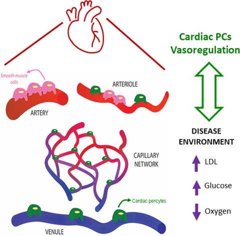
Cardiac pericytes function as key vasoactive cells to regulate homeostasis and disease
FEBS Open Bio ( IF 2.8 ) Pub Date : 2020-11-01 , DOI: 10.1002/2211-5463.13021 Linda L Lee 1 , Aarif Y Khakoo 2 , Vishnu Chintalgattu 1
FEBS Open Bio ( IF 2.8 ) Pub Date : 2020-11-01 , DOI: 10.1002/2211-5463.13021 Linda L Lee 1 , Aarif Y Khakoo 2 , Vishnu Chintalgattu 1
Affiliation
|
Pericytes (PCs)—mural cells that envelop endothelial cells (ECs) of microvessels—regulate tissue‐specific vasculature development as well as maturation and maintenance of endothelial barrier integrity. However, little is known about their tissue‐specific function in the heart. Specifically, the mechanism by which cardiac PCs constrict coronary capillaries remains undetermined. To gain insights into the function of cardiac PCs at the cellular level, we isolated NG2+ PDGFRβ+ CD146+ CD34− CD31− CD45− PCs for detailed characterization. Functionally, we provide evidence that these PCs increased transepithelial electrical resistance and decreased endothelial permeability. We show for the first time that this population of PCs express contractile proteins, are stimulated by adrenergic signaling, and demonstrate stereotypical contraction and relaxation. Furthermore, we also studied for the first time, the PCs in in vitro models of disease. PCs in hypoxia activated the hypoxia‐inducible factor 1 alpha pathway, increased secretion of angiogenic factors, and caused cellular apoptosis. Supraphysiological levels of low‐density lipoprotein decreased PC proliferation and induced lipid droplet accumulation. Elevated glucose levels triggered a proinflammatory response. Taken together, our study characterizes cardiac PCs under in vitro disease conditions and supports the hypothesis that cardiac PCs are key vasoactive cells that can regulate blood flow in the heart.
中文翻译:

心脏周细胞是调节体内平衡和疾病的关键血管活性细胞
周细胞 (PCs) - 包裹微血管内皮细胞 (ECs) 的壁细胞 - 调节组织特异性脉管系统发育以及内皮屏障完整性的成熟和维持。然而,关于它们在心脏中的组织特异性功能知之甚少。具体而言,心脏 PC 收缩冠状毛细血管的机制仍未确定。为了在细胞水平上深入了解心脏 PC 的功能,我们分离了 NG2 + PDGFRβ + CD146 + CD34 - CD31 - CD45 -用于详细表征的 PC。在功能上,我们提供证据表明这些 PC 增加了跨上皮电阻并降低了内皮通透性。我们首次表明,这群个人电脑表达收缩蛋白,受到肾上腺素能信号的刺激,并表现出典型的收缩和放松。此外,我们还首次研究了体外PC疾病模型。缺氧状态下的 PC 激活了缺氧诱导因子 1 α 通路,增加了血管生成因子的分泌,并导致细胞凋亡。超生理水平的低密度脂蛋白降低了 PC 增殖并诱导了脂滴积累。葡萄糖水平升高引发促炎反应。总之,我们的研究表征了体外疾病条件下的心脏 PC,并支持以下假设:心脏 PC 是可以调节心脏血流的关键血管活性细胞。
更新日期:2021-01-04
中文翻译:
心脏周细胞是调节体内平衡和疾病的关键血管活性细胞
周细胞 (PCs) - 包裹微血管内皮细胞 (ECs) 的壁细胞 - 调节组织特异性脉管系统发育以及内皮屏障完整性的成熟和维持。然而,关于它们在心脏中的组织特异性功能知之甚少。具体而言,心脏 PC 收缩冠状毛细血管的机制仍未确定。为了在细胞水平上深入了解心脏 PC 的功能,我们分离了 NG2 + PDGFRβ + CD146 + CD34 - CD31 - CD45 -用于详细表征的 PC。在功能上,我们提供证据表明这些 PC 增加了跨上皮电阻并降低了内皮通透性。我们首次表明,这群个人电脑表达收缩蛋白,受到肾上腺素能信号的刺激,并表现出典型的收缩和放松。此外,我们还首次研究了体外PC疾病模型。缺氧状态下的 PC 激活了缺氧诱导因子 1 α 通路,增加了血管生成因子的分泌,并导致细胞凋亡。超生理水平的低密度脂蛋白降低了 PC 增殖并诱导了脂滴积累。葡萄糖水平升高引发促炎反应。总之,我们的研究表征了体外疾病条件下的心脏 PC,并支持以下假设:心脏 PC 是可以调节心脏血流的关键血管活性细胞。


 京公网安备 11010802027423号