当前位置:
X-MOL 学术
›
J. Biophotonics
›
论文详情
Our official English website, www.x-mol.net, welcomes your feedback! (Note: you will need to create a separate account there.)
Microscopic phase reconstruction of cervical exfoliated cell under partially coherent illumination
Journal of Biophotonics ( IF 2.8 ) Pub Date : 2020-10-31 , DOI: 10.1002/jbio.202000401 Xingyuan Lu 1 , Zhuoyi Wang 1 , Suxia Zhang 2 , A P Konijnenberg 3 , Yiqin Ouyang 2 , Chengliang Zhao 1 , Yangjian Cai 1, 4, 5
Journal of Biophotonics ( IF 2.8 ) Pub Date : 2020-10-31 , DOI: 10.1002/jbio.202000401 Xingyuan Lu 1 , Zhuoyi Wang 1 , Suxia Zhang 2 , A P Konijnenberg 3 , Yiqin Ouyang 2 , Chengliang Zhao 1 , Yangjian Cai 1, 4, 5
Affiliation
|
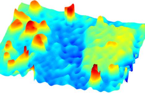
Basic coherent diffraction imaging methods strongly rely on having a highly coherent illumination in order to reconstruct the phase accurately. However, regardless of considering the turbulent transport medium, the instability of the system or the generation mechanism of the light source, partially coherent illumination is more common in real case. In this paper, we proposed an efficient microscopic phase imaging method to study normal and abnormal cervical exfoliated cells. By applying three phase modulations in a single point of the sample's transmitted field, the phase can be retrieved with correspoding three intensities under partially coherent illumination. Compared with intensity map, we can efficiently and clearly judge the proportion of high density shrinking abnormal cells from the phase distributions, which provides a confident analysis and evaluation basis for early medical diagnosis of cervical cancer. This study also has potential applications in noninvasive optical imaging of dynamic biological tissues.
中文翻译:

部分相干照明下宫颈脱落细胞的显微相重建
基本相干衍射成像方法强烈依赖于具有高度相干的照明,以便准确地重建相位。但是,无论考虑湍流的输送介质,系统的不稳定性还是光源的产生机理,在实际情况下,局部相干照明更为常见。在本文中,我们提出了一种有效的显微相成像方法来研究正常和异常的宫颈脱落细胞。通过在样品的透射场的单个点上应用三相调制,可以在部分相干照明下以对应的三个强度来恢复相位。与强度图相比,我们可以从相分布中高效,清晰地判断高密度收缩异常细胞的比例,为宫颈癌的早期医学诊断提供了可靠的分析和评价依据。这项研究在动态生物组织的无创光学成像中也具有潜在的应用。
更新日期:2021-01-04
中文翻译:
部分相干照明下宫颈脱落细胞的显微相重建
基本相干衍射成像方法强烈依赖于具有高度相干的照明,以便准确地重建相位。但是,无论考虑湍流的输送介质,系统的不稳定性还是光源的产生机理,在实际情况下,局部相干照明更为常见。在本文中,我们提出了一种有效的显微相成像方法来研究正常和异常的宫颈脱落细胞。通过在样品的透射场的单个点上应用三相调制,可以在部分相干照明下以对应的三个强度来恢复相位。与强度图相比,我们可以从相分布中高效,清晰地判断高密度收缩异常细胞的比例,为宫颈癌的早期医学诊断提供了可靠的分析和评价依据。这项研究在动态生物组织的无创光学成像中也具有潜在的应用。


 京公网安备 11010802027423号