当前位置:
X-MOL 学术
›
Photochem. Photobiol.
›
论文详情
Our official English website, www.x-mol.net, welcomes your
feedback! (Note: you will need to create a separate account there.)
Painless Photodynamic Therapy Triggers Innate and Adaptive Immune Responses in a Murine Model of UV‐induced Squamous Skin Pre‐cancer
Photochemistry and Photobiology ( IF 2.6 ) Pub Date : 2020-11-22 , DOI: 10.1111/php.13350 Sanjay Anand 1, 2, 3 , Mukul Govande 1 , Anton Yasinchak 1 , Lauren Heusinkveld 3 , Sajina Shakya 1 , Robert L Fairchild 3, 4 , Edward V Maytin 1, 2, 3
Photochemistry and Photobiology ( IF 2.6 ) Pub Date : 2020-11-22 , DOI: 10.1111/php.13350 Sanjay Anand 1, 2, 3 , Mukul Govande 1 , Anton Yasinchak 1 , Lauren Heusinkveld 3 , Sajina Shakya 1 , Robert L Fairchild 3, 4 , Edward V Maytin 1, 2, 3
Affiliation
|
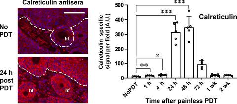
Painless photodynamic therapy (p-PDT), which involves application of photosensitizer and immediate exposure to light to treat actinic keratosis (AK) in patients, causes negligible pain on the day of treatment but leads to delayed inflammation and effective lesion clearance (Kaw et al, J Am Acad Dermatol 2020). To better understand how p-PDT works, hairless mice with UV-induced AK were treated with p-PDT and monitored for 2 weeks. Lesion clearance after p-PDT was similar to clearance after conventional PDT (c-PDT). However, lesion biopsies showed minimal cell death and less production of reactive oxygen species (ROS) in p-PDT-treated than in c-PDT-treated lesions. Interestingly, p-PDT triggered vigorous recruitment of immune cells associated with innate immunity. Neutrophils (Ly6G+) and macrophages (F4/80+) appeared at 4 h and peaked at 24 h after p-PDT. Damage Associated Molecular Patterns (DAMPs) including calreticulin, HMGB1, and HSP70, were expressed at maximum levels around 24 h post p-PDT. Total T-cells (CD3+) were increased at 24 h, whereas large increases in cytotoxic (CD8+) and regulatory (Foxp3+) T-cells were observed at 1- and 2- weeks post p-PDT. In summary, the ability of p-PDT to eliminate AK lesions, despite very little overt cellular damage, appears to involve stimulation of a local immune response.
中文翻译:

无痛光动力疗法在紫外线诱发的鳞状皮肤癌前期小鼠模型中触发先天性和适应性免疫反应
无痛光动力疗法 (p-PDT) 涉及应用光敏剂并立即接受光线治疗患者的光化性角化病 (AK),治疗当天的疼痛可以忽略不计,但会延迟炎症并有效清除病灶(Kaw 等人) ,《J Am Acad Dermatol》2020)。为了更好地了解 p-PDT 的工作原理,对患有紫外线诱导 AK 的无毛小鼠进行 p-PDT 治疗并监测 2 周。p-PDT 后的病灶清除率与传统 PDT (c-PDT) 后的清除率相似。然而,病灶活检显示,与 c-PDT 治疗的病灶相比,p-PDT 治疗的病灶中细胞死亡最少,活性氧 (ROS) 的产生也更少。有趣的是,p-PDT 引发了与先天免疫相关的免疫细胞的活跃募集。中性粒细胞 (Ly6G+) 和巨噬细胞 (F4/80+) 在 p-PDT 后 4 小时出现,并在 24 小时达到峰值。损伤相关分子模式 (DAMP) 包括钙网蛋白、HMGB1 和 HSP70,在 p-PDT 后 24 小时左右表达达到最高水平。总 T 细胞 (CD3+) 在 24 小时增加,而细胞毒性 (CD8+) 和调节性 (Foxp3+) T 细胞在 p-PDT 后 1 周和 2 周大幅增加。总之,p-PDT 消除 AK 病变的能力,尽管明显的细胞损伤很少,但似乎涉及刺激局部免疫反应。
更新日期:2020-11-22
中文翻译:
无痛光动力疗法在紫外线诱发的鳞状皮肤癌前期小鼠模型中触发先天性和适应性免疫反应
无痛光动力疗法 (p-PDT) 涉及应用光敏剂并立即接受光线治疗患者的光化性角化病 (AK),治疗当天的疼痛可以忽略不计,但会延迟炎症并有效清除病灶(Kaw 等人) ,《J Am Acad Dermatol》2020)。为了更好地了解 p-PDT 的工作原理,对患有紫外线诱导 AK 的无毛小鼠进行 p-PDT 治疗并监测 2 周。p-PDT 后的病灶清除率与传统 PDT (c-PDT) 后的清除率相似。然而,病灶活检显示,与 c-PDT 治疗的病灶相比,p-PDT 治疗的病灶中细胞死亡最少,活性氧 (ROS) 的产生也更少。有趣的是,p-PDT 引发了与先天免疫相关的免疫细胞的活跃募集。中性粒细胞 (Ly6G+) 和巨噬细胞 (F4/80+) 在 p-PDT 后 4 小时出现,并在 24 小时达到峰值。损伤相关分子模式 (DAMP) 包括钙网蛋白、HMGB1 和 HSP70,在 p-PDT 后 24 小时左右表达达到最高水平。总 T 细胞 (CD3+) 在 24 小时增加,而细胞毒性 (CD8+) 和调节性 (Foxp3+) T 细胞在 p-PDT 后 1 周和 2 周大幅增加。总之,p-PDT 消除 AK 病变的能力,尽管明显的细胞损伤很少,但似乎涉及刺激局部免疫反应。


 京公网安备 11010802027423号