当前位置:
X-MOL 学术
›
J. Raman Spectrosc.
›
论文详情
Our official English website, www.x-mol.net, welcomes your
feedback! (Note: you will need to create a separate account there.)
Principal component analysis facilitated fast and noninvasive Raman spectroscopic imaging of plant cell wall pectin distribution and interaction with enzymatic hydrolysis
Journal of Raman Spectroscopy ( IF 2.4 ) Pub Date : 2020-10-28 , DOI: 10.1002/jrs.6022 Qing He 1 , Olga A. Zabotina 2 , Chenxu Yu 1
Journal of Raman Spectroscopy ( IF 2.4 ) Pub Date : 2020-10-28 , DOI: 10.1002/jrs.6022 Qing He 1 , Olga A. Zabotina 2 , Chenxu Yu 1
Affiliation
|
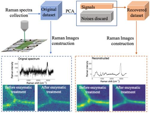
The plant cell wall is a complex network of polysaccharides. A better understanding of the plant cell wall polysaccharide content, distribution, and interactions among them could be instrumental for our further understanding of plant cell wall in general. Confocal Raman microscopy (CRM) is a powerful tool that could reveal details about chemical landscape of the plant cell wall with micrometer resolution. However, the low signal‐to‐noise (S/N) ratio of Raman spectral signal led to low throughput of Raman imaging, which limited its application in plant cell wall study. In this study, the interaction between the cell wall polysaccharides and the pectin enzyme endo‐polygalacturonase (EPG) was characterized by analyzing Raman images collected before and after EPG treatment at high scanning speed. The obtained low S/N ratio Raman spectra were processed with principal component analysis (PCA) and hierarchical clustering analysis (HCA) to recover information‐rich signature changes in Raman signal dataset that reflected the changes in the polysaccharides caused specifically by the enzymatic hydrolysis. The PCA reconstruction technique significantly improved the S/N ratio of the spectra dataset while kept the Raman signal intact. Further analysis of principal components (PCs) revealed the pectin distribution and its interaction with the enzyme, which provided organizational details of pectin inside the onion plant cell wall. The technique could be used to reveal polysaccharide organization and distribution inside plant cell walls.
中文翻译:

主成分分析促进了植物细胞壁果胶分布以及与酶水解相互作用的快速无创拉曼光谱成像
植物细胞壁是多糖的复杂网络。更好地了解植物细胞壁多糖的含量,分布及其之间的相互作用可能有助于我们进一步了解植物细胞壁。共焦拉曼显微镜(CRM)是一种功能强大的工具,可以用千分尺分辨率揭示有关植物细胞壁化学景观的细节。然而,拉曼光谱信号的低信噪比导致低通量的拉曼成像,这限制了其在植物细胞壁研究中的应用。在这项研究中,通过分析在EPG处理之前和之后以高扫描速度采集的拉曼图像,来表征细胞壁多糖与果胶酶内聚半乳糖醛酸酶(EPG)之间的相互作用。使用主成分分析(PCA)和层次聚类分析(HCA)处理获得的低信噪比拉曼光谱,以恢复拉曼信号数据集中信息丰富的特征变化,这些变化反映了酶水解特别引起的多糖变化。PCA重建技术显着提高了光谱数据集的信噪比,同时保持了拉曼信号的完整性。对主要成分(PCs)的进一步分析揭示了果胶的分布及其与酶的相互作用,从而提供了洋葱植物细胞壁内果胶的组织细节。该技术可用于揭示植物细胞壁内多糖的组织和分布。
更新日期:2020-12-10
中文翻译:
主成分分析促进了植物细胞壁果胶分布以及与酶水解相互作用的快速无创拉曼光谱成像
植物细胞壁是多糖的复杂网络。更好地了解植物细胞壁多糖的含量,分布及其之间的相互作用可能有助于我们进一步了解植物细胞壁。共焦拉曼显微镜(CRM)是一种功能强大的工具,可以用千分尺分辨率揭示有关植物细胞壁化学景观的细节。然而,拉曼光谱信号的低信噪比导致低通量的拉曼成像,这限制了其在植物细胞壁研究中的应用。在这项研究中,通过分析在EPG处理之前和之后以高扫描速度采集的拉曼图像,来表征细胞壁多糖与果胶酶内聚半乳糖醛酸酶(EPG)之间的相互作用。使用主成分分析(PCA)和层次聚类分析(HCA)处理获得的低信噪比拉曼光谱,以恢复拉曼信号数据集中信息丰富的特征变化,这些变化反映了酶水解特别引起的多糖变化。PCA重建技术显着提高了光谱数据集的信噪比,同时保持了拉曼信号的完整性。对主要成分(PCs)的进一步分析揭示了果胶的分布及其与酶的相互作用,从而提供了洋葱植物细胞壁内果胶的组织细节。该技术可用于揭示植物细胞壁内多糖的组织和分布。


 京公网安备 11010802027423号