Journal of Structural Biology ( IF 3.0 ) Pub Date : 2020-10-24 , DOI: 10.1016/j.jsb.2020.107659 Inna Bukreeva 1 , Olga Junemann 2 , Alessia Cedola 3 , Francesca Palermo 4 , Laura Maugeri 5 , Ginevra Begani Provinciali 6 , Nicola Pieroni 7 , Alessia Sanna 3 , Dmitry A Otlyga 2 , Alexey Buzmakov 8 , Yuri Krivonosov 8 , Denis Zolotov 8 , Marina Chukalina 9 , Anna Ivanova 8 , Sergey Saveliev 2 , Victor Asadchikov 8 , Michela Fratini 5
|
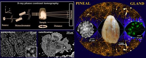
Pineal gland (PG) is a part of the human brain epithalamus that plays an important role in sleep, circadian rhythm, immunity, and reproduction. The calcium deposits and lesions in PG interfere with normal function of the organ and can be associated with different health disorders including serious neurological diseases. At the moment, the detailed mechanisms of PG calcifications and PG lesions formation as well as their involvement in pathological processes are not fully understood. The deep and comprehensive study of the structure of the uncut human PG with histological details, poses a stiff challenge to most imaging techniques, due to low spatial resolution, low visibility or to exceedingly aggressive sample preparation. Here, we investigate the whole uncut and unstained human post-mortem PGs by X-ray phase contrast tomography (XPCT). XPCT is an advanced 3D imaging technique, that permits to study of both soft and calcified tissue of a sample at different scales: from the whole organ to cell structure. In our research we simultaneously resolved 3D structure of parenchyma, vascular network and calcifications. Moreover, we distinguished structural details of intact and degenerated PG tissue. We discriminated calcifications with different structure, pinealocytes nuclei and the glial cells processes. All results were validated by histology. Our research clear demonstrated that XPCT is a potential tool for the high resolution 3D imaging of PG morphological features. This technique opens a new perspective to investigate PG dysfunction and understand the mechanisms of onset and progression of diseases involving the pineal gland.
中文翻译:
通过 X 射线相衬断层扫描对人体松果体 3D 组织的研究
松果体 (PG) 是人脑上丘脑的一部分,在睡眠、昼夜节律、免疫和繁殖中起着重要作用。PG 中的钙沉积和病变会干扰器官的正常功能,并可能与包括严重神经系统疾病在内的不同健康障碍有关。目前,PG钙化和PG病变形成的详细机制及其参与病理过程尚不完全清楚。由于空间分辨率低、能见度低或样品制备过于激进,因此对具有组织学细节的未切割人类 PG 结构进行深入而全面的研究,对大多数成像技术提出了严峻的挑战。在这里,我们通过 X 射线相衬断层扫描 (XPCT) 调查整个未切割和未染色的人类死后 PG。XPCT 是一种先进的 3D 成像技术,可以在不同尺度上研究样本的软组织和钙化组织:从整个器官到细胞结构。在我们的研究中,我们同时解析了实质、血管网络和钙化的 3D 结构。此外,我们区分了完整和退化的 PG 组织的结构细节。我们区分了不同结构的钙化、松果体细胞核和神经胶质细胞过程。所有结果均经组织学验证。我们的研究清楚地表明,XPCT 是对 PG 形态特征进行高分辨率 3D 成像的潜在工具。该技术为研究 PG 功能障碍和了解涉及松果体的疾病的发生和进展机制开辟了新的视角。这允许在不同尺度上研究样本的软组织和钙化组织:从整个器官到细胞结构。在我们的研究中,我们同时解析了实质、血管网络和钙化的 3D 结构。此外,我们区分了完整和退化的 PG 组织的结构细节。我们区分了不同结构的钙化、松果体细胞核和神经胶质细胞过程。所有结果均经组织学验证。我们的研究清楚地表明,XPCT 是对 PG 形态特征进行高分辨率 3D 成像的潜在工具。该技术为研究 PG 功能障碍和了解涉及松果体的疾病的发生和进展机制开辟了新的视角。这允许在不同尺度上研究样本的软组织和钙化组织:从整个器官到细胞结构。在我们的研究中,我们同时解析了实质、血管网络和钙化的 3D 结构。此外,我们区分了完整和退化的 PG 组织的结构细节。我们区分了不同结构的钙化、松果体细胞核和神经胶质细胞过程。所有结果均经组织学验证。我们的研究清楚地表明,XPCT 是对 PG 形态特征进行高分辨率 3D 成像的潜在工具。该技术为研究 PG 功能障碍和了解涉及松果体的疾病的发生和进展机制开辟了新的视角。从整个器官到细胞结构。在我们的研究中,我们同时解析了实质、血管网络和钙化的 3D 结构。此外,我们区分了完整和退化的 PG 组织的结构细节。我们区分了不同结构的钙化、松果体细胞核和神经胶质细胞过程。所有结果均经组织学验证。我们的研究清楚地表明,XPCT 是对 PG 形态特征进行高分辨率 3D 成像的潜在工具。该技术为研究 PG 功能障碍和了解涉及松果体的疾病的发生和进展机制开辟了新的视角。从整个器官到细胞结构。在我们的研究中,我们同时解析了实质、血管网络和钙化的 3D 结构。此外,我们区分了完整和退化的 PG 组织的结构细节。我们区分了不同结构的钙化、松果体细胞核和神经胶质细胞过程。所有结果均通过组织学验证。我们的研究清楚地表明,XPCT 是对 PG 形态特征进行高分辨率 3D 成像的潜在工具。该技术为研究 PG 功能障碍和了解涉及松果体的疾病的发生和进展机制开辟了新的视角。我们区分了完整和退化的 PG 组织的结构细节。我们区分了不同结构的钙化、松果体细胞核和神经胶质细胞过程。所有结果均经组织学验证。我们的研究清楚地表明,XPCT 是对 PG 形态特征进行高分辨率 3D 成像的潜在工具。该技术为研究 PG 功能障碍和了解涉及松果体的疾病的发生和进展机制开辟了新的视角。我们区分了完整和退化的 PG 组织的结构细节。我们区分了不同结构的钙化、松果体细胞核和神经胶质细胞过程。所有结果均经组织学验证。我们的研究清楚地表明,XPCT 是对 PG 形态特征进行高分辨率 3D 成像的潜在工具。该技术为研究 PG 功能障碍和了解涉及松果体的疾病的发生和进展机制开辟了新的视角。


 京公网安备 11010802027423号