Journal of Structural Biology ( IF 3.0 ) Pub Date : 2020-10-23 , DOI: 10.1016/j.jsb.2020.107653 Gaia Crippa 1 , Erika Griesshaber 2 , Antonio G Checa 3 , Elizabeth M Harper 4 , Maria Simonet Roda 2 , Wolfgang W Schmahl 2
|
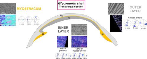
The shells of the bivalves Glycymeris glycymeris and Glycymeris nummaria are widely used for environmental studies. They consist of aragonite and comprise four different microstructures and textures from outer to inner shell surfaces: crossed-lamellar, myostracal, complex crossed-lamellar and fibrous prismatic. We characterize with SEM, EBSD, laser-confocal microscopy and AFM imaging mineral unit size, morphology and orientation of crystallites in the different microstructural arrangements and at the transition from one microstructure to the other. We also characterize the microstructure and texture of adductor and pedal retractor myostraca and address structural characteristics at the transition from crossed-lamellar to myostracal assemblies. We find that the crossed-lamellar layer has a three-dimensional crystallographic orientational order. Each set of first-order lamellae consists of twinned aragonite; the two sets of first-order lamellae are misoriented to each other by about 30 to 40° while retaining an approximately parallel a-axis; they do not show any particular twin relationship. Myostracal aragonite grows homoepitactically onto the crossed-lamellar aragonite, but is clearly a separate microstructure, with its own crystallite size and morphology. Within adductor and pedal myostraca, prisms increase in size towards inner surfaces. In contrast to the other shell layers, the myostraca form through competitive growth. The complex crossed-lamellar aragonite initially inherits the three-dimensional texture of the crossed-lamellar microstructure, but with growth develops an axial texture, which is transmitted to the underlying fibrous prismatic microstructure. With this work we provide a modern, unaltered, reference for fossil Glycymeris shells to be used for detection of diagenetic overprint in fossil Glycymeris analogs.
中文翻译:
现代甘草壳的文石交叉层状、纤维棱柱状和肌层微结构的取向模式
双壳类Glycymeris glycymeris和Glycymeris nummaria的壳广泛用于环境研究。它们由文石组成,包括四种不同的微观结构和从外壳表面到内壳表面的纹理:交叉层状、肌层状、复杂交叉层状和纤维棱柱状。我们用 SEM、EBSD、激光共聚焦显微镜和 AFM 成像矿物单元大小、形态和微晶在不同微观结构排列中的取向以及从一种微观结构到另一种微观结构的转变进行表征。我们还表征了内收肌和踏板牵开器 myostraca 的微观结构和质地,并解决了从交叉层状组件到 myostracal 组件过渡时的结构特征。我们发现交叉层状层具有三维晶体取向顺序。每组一级片晶由孪晶文石组成;两组一阶薄片彼此错位约 30 至 40°,同时保持大致平行的 a 轴;他们没有表现出任何特殊的孪生关系。Myostracal 文石同晶生长在交叉层状文石上,但显然是一种独立的微观结构,具有自己的微晶尺寸和形态。在内收肌和足肌内,棱柱朝向内表面增大。与其他壳层相反,myostraca 通过竞争性生长形成。复杂的交叉层状文石最初继承了交叉层状微观结构的三维织构,但随着生长形成轴向织构,并传递到下面的纤维棱柱状微观结构。通过这项工作,我们为化石提供了一个现代的、不变的参考 他们没有表现出任何特殊的孪生关系。Myostracal 文石在交叉层状文石上同晶生长,但显然是一种独立的微观结构,具有自己的微晶尺寸和形态。在内收肌和足肌内,棱柱朝向内表面增大。与其他壳层相反,myostraca 通过竞争性生长形成。复杂的交叉层状文石最初继承了交叉层状微观结构的三维织构,但随着增长发展出轴向织构,并传递到下面的纤维棱柱状微观结构。通过这项工作,我们为化石提供了一个现代的、不变的参考 他们没有表现出任何特殊的孪生关系。Myostracal 文石同晶生长在交叉层状文石上,但显然是一种独立的微观结构,具有自己的微晶尺寸和形态。在内收肌和足肌内,棱柱朝向内表面增大。与其他壳层相比,myostraca 通过竞争性生长形成。复杂的交叉层状文石最初继承了交叉层状微观结构的三维织构,但随着增长发展出轴向织构,并传递到下面的纤维棱柱状微观结构。通过这项工作,我们为化石提供了一个现代的、不变的参考 具有自己的微晶尺寸和形态。在内收肌和足肌内,棱柱朝向内表面增大。与其他壳层相反,myostraca 通过竞争性生长形成。复杂的交叉层状文石最初继承了交叉层状微观结构的三维织构,但随着增长发展出轴向织构,并传递到下面的纤维棱柱状微观结构。通过这项工作,我们为化石提供了一个现代的、不变的参考 具有自己的微晶尺寸和形态。在内收肌和足肌内,棱柱朝向内表面的尺寸增加。与其他壳层相反,myostraca 通过竞争性生长形成。复杂的交叉层状文石最初继承了交叉层状微观结构的三维织构,但随着增长发展出轴向织构,并传递到下面的纤维棱柱状微观结构。通过这项工作,我们为化石提供了一个现代的、不变的参考 但随着生长会形成轴向纹理,并传递到下面的纤维棱柱微结构。通过这项工作,我们为化石提供了一个现代的、不变的参考 但随着生长会形成轴向纹理,并传递到下面的纤维棱柱微结构。通过这项工作,我们为化石提供了一个现代的、不变的参考用于检测Glycymeris化石类似物中成岩叠印的Glycymeris壳。


 京公网安备 11010802027423号