Developmental Cell ( IF 10.7 ) Pub Date : 2020-10-21 , DOI: 10.1016/j.devcel.2020.09.030 Claire S Simon 1 , Shahadat Rahman 1 , Dhruv Raina 2 , Christian Schröter 2 , Anna-Katerina Hadjantonakis 1
|
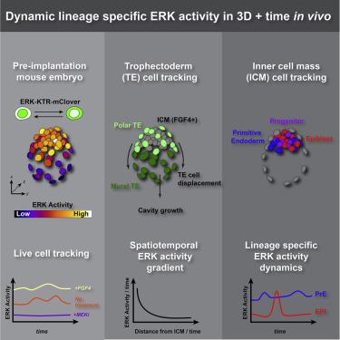
FGF/ERK signaling is crucial for the patterning and proliferation of cell lineages that comprise the mouse blastocyst. However, ERK signaling dynamics have never been directly visualized in live embryos. To address whether differential signaling is associated with particular cell fates and states, we generated a targeted mouse line expressing an ERK-kinase translocation reporter (KTR) that enables live quantification of ERK activity at single-cell resolution. 3D time-lapse imaging of this biosensor in embryos revealed spatially graded ERK activity in the trophectoderm prior to overt polar versus mural differentiation. Within the inner cell mass (ICM), all cells relayed FGF/ERK signals with varying durations and magnitude. Primitive endoderm cells displayed higher overall levels of ERK activity, while pluripotent epiblast cells exhibited lower basal activity with sporadic pulses. These results constitute a direct visualization of signaling events during mammalian pre-implantation development and reveal the existence of spatial and temporal lineage-specific dynamics.
中文翻译:
小鼠囊胚中 ERK 活性的实时可视化揭示了谱系特异性信号传导动力学
FGF/ERK 信号传导对于构成小鼠囊胚的细胞谱系的模式化和增殖至关重要。然而,ERK 信号动力学从未在活胚胎中直接观察到。为了解决差异信号传导是否与特定细胞命运和状态相关的问题,我们生成了表达 ERK 激酶易位报告基因 (KTR) 的靶向小鼠品系,该小鼠品系能够以单细胞分辨率实时定量 ERK 活性。该生物传感器在胚胎中的 3D 延时成像揭示了在明显的极性分化和壁分化之前滋养外胚层中 ERK 活性的空间分级。在内细胞团 (ICM) 内,所有细胞都以不同的持续时间和强度传递 FGF/ERK 信号。原始内胚层细胞表现出较高的 ERK 活性总体水平,而多能外胚层细胞则表现出较低的基础活性和零星脉冲。这些结果构成了哺乳动物植入前发育过程中信号事件的直接可视化,并揭示了空间和时间谱系特异性动态的存在。


 京公网安备 11010802027423号