当前位置:
X-MOL 学术
›
Prog. Nat. Sci. Mater. Int.
›
论文详情
Our official English website, www.x-mol.net, welcomes your
feedback! (Note: you will need to create a separate account there.)
Patterning the neuronal cells via inkjet printing of self-assembled peptides on silk scaffolds
Progress in Natural Science: Materials International ( IF 4.8 ) Pub Date : 2020-10-01 , DOI: 10.1016/j.pnsc.2020.09.007 Weizhen Sun , Yi Zhang , David A. Gregory , Ana Jimenez-Franco , Mhd Anas Tomeh , Songwei Lv , Jiqian Wang , John W. Haycock , Jian R. Lu , Xiubo Zhao
Progress in Natural Science: Materials International ( IF 4.8 ) Pub Date : 2020-10-01 , DOI: 10.1016/j.pnsc.2020.09.007 Weizhen Sun , Yi Zhang , David A. Gregory , Ana Jimenez-Franco , Mhd Anas Tomeh , Songwei Lv , Jiqian Wang , John W. Haycock , Jian R. Lu , Xiubo Zhao
|
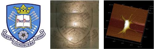
Abstract The patterning of neuronal cells and guiding neurite growth are important for neuron tissue engineering and cell-based biosensors. In this paper, inkjet printing has been employed to pattern self-assembled I3QGK peptide nanofibers on silk substrates for guiding the growth of neuron-like PC12 cells. Atomic force microscopy (AFM) confirmed the dynamic self-assembly of I3QGK into nanofiber structures. The printed self-assembled peptide strongly adheres to regenerated silk fibroin (RSF) substrates through charge-charge interactions. It was observed that in the absence of I3QGK, PC12 cells exhibited poor attachment to RSF films, while for RSF surfaces coated or printed with peptide nanofibers, cellular attachment was significantly improved in terms of both cell density and morphology. AFM results revealed that peptide nanofibers can promote the generation of axons and terminal buttons of PC12 cells, indicating that I3QGK nanofibers not only promote cellular attachment but also facilitate differentiation into neuronal phenotypes. Inkjet printing allows complex patterning of peptide nanofibers onto RSF substrates, which enabled us to engineer cell alignment and provide an opportunity to direct axonal development in vitro. The live/dead assay showed that printed I3QGK patterns exhibit no cytotoxicity to PC12 cells demonstrating potential for future nerve tissue engineering applications.
中文翻译:

通过在丝支架上喷墨打印自组装肽来对神经元细胞进行图案化
摘要 神经元细胞的模式化和指导神经突生长对于神经元组织工程和基于细胞的生物传感器很重要。在本文中,喷墨印刷已被用于在丝绸基材上对自组装的 I3QGK 肽纳米纤维进行图案化,以指导神经元样 PC12 细胞的生长。原子力显微镜 (AFM) 证实了 I3QGK 动态自组装成纳米纤维结构。打印的自组装肽通过电荷-电荷相互作用强烈粘附在再生丝素蛋白 (RSF) 基材上。观察到在不存在 I3QGK 的情况下,PC12 细胞对 RSF 膜的附着力较差,而对于用肽纳米纤维涂覆或印刷的 RSF 表面,细胞密度和形态方面的细胞附着均显着改善。AFM 结果显示,肽纳米纤维可以促进 PC12 细胞轴突和末端按钮的产生,表明 I3QGK 纳米纤维不仅促进细胞附着,还促进向神经元表型的分化。喷墨打印允许在 RSF 基底上形成复杂的肽纳米纤维图案,这使我们能够设计细胞排列并提供在体外指导轴突发育的机会。活/死试验表明,打印的 I3QGK 模式对 PC12 细胞没有细胞毒性,这表明在未来的神经组织工程应用中具有潜力。喷墨打印允许在 RSF 基底上形成复杂的肽纳米纤维图案,这使我们能够设计细胞排列并提供在体外指导轴突发育的机会。活/死试验表明,打印的 I3QGK 模式对 PC12 细胞没有细胞毒性,这表明在未来的神经组织工程应用中具有潜力。喷墨打印允许在 RSF 基底上形成复杂的肽纳米纤维图案,这使我们能够设计细胞排列并提供在体外指导轴突发育的机会。活/死试验表明,打印的 I3QGK 模式对 PC12 细胞没有细胞毒性,这表明在未来的神经组织工程应用中具有潜力。
更新日期:2020-10-01
中文翻译:
通过在丝支架上喷墨打印自组装肽来对神经元细胞进行图案化
摘要 神经元细胞的模式化和指导神经突生长对于神经元组织工程和基于细胞的生物传感器很重要。在本文中,喷墨印刷已被用于在丝绸基材上对自组装的 I3QGK 肽纳米纤维进行图案化,以指导神经元样 PC12 细胞的生长。原子力显微镜 (AFM) 证实了 I3QGK 动态自组装成纳米纤维结构。打印的自组装肽通过电荷-电荷相互作用强烈粘附在再生丝素蛋白 (RSF) 基材上。观察到在不存在 I3QGK 的情况下,PC12 细胞对 RSF 膜的附着力较差,而对于用肽纳米纤维涂覆或印刷的 RSF 表面,细胞密度和形态方面的细胞附着均显着改善。AFM 结果显示,肽纳米纤维可以促进 PC12 细胞轴突和末端按钮的产生,表明 I3QGK 纳米纤维不仅促进细胞附着,还促进向神经元表型的分化。喷墨打印允许在 RSF 基底上形成复杂的肽纳米纤维图案,这使我们能够设计细胞排列并提供在体外指导轴突发育的机会。活/死试验表明,打印的 I3QGK 模式对 PC12 细胞没有细胞毒性,这表明在未来的神经组织工程应用中具有潜力。喷墨打印允许在 RSF 基底上形成复杂的肽纳米纤维图案,这使我们能够设计细胞排列并提供在体外指导轴突发育的机会。活/死试验表明,打印的 I3QGK 模式对 PC12 细胞没有细胞毒性,这表明在未来的神经组织工程应用中具有潜力。喷墨打印允许在 RSF 基底上形成复杂的肽纳米纤维图案,这使我们能够设计细胞排列并提供在体外指导轴突发育的机会。活/死试验表明,打印的 I3QGK 模式对 PC12 细胞没有细胞毒性,这表明在未来的神经组织工程应用中具有潜力。


 京公网安备 11010802027423号