当前位置:
X-MOL 学术
›
Eur. J. Immunol.
›
论文详情
Our official English website, www.x-mol.net, welcomes your
feedback! (Note: you will need to create a separate account there.)
Plasma membrane Ca2+ ATPase 1 (PMCA1) but not PMCA4 is critical for B‐cell development and Ca2+ homeostasis in mice
European Journal of Immunology ( IF 4.5 ) Pub Date : 2020-10-24 , DOI: 10.1002/eji.202048654 Mark Korthals 1 , Laura Tech 1 , Kristina Langnaese 1 , Anna Gottfried 1 , Johannes Hradsky 1 , Ulrich Thomas 2 , Ana Claudia Zenclussen 3 , Monika C Brunner-Weinzierl 4 , Kerry Tedford 1 , Klaus-Dieter Fischer 1
European Journal of Immunology ( IF 4.5 ) Pub Date : 2020-10-24 , DOI: 10.1002/eji.202048654 Mark Korthals 1 , Laura Tech 1 , Kristina Langnaese 1 , Anna Gottfried 1 , Johannes Hradsky 1 , Ulrich Thomas 2 , Ana Claudia Zenclussen 3 , Monika C Brunner-Weinzierl 4 , Kerry Tedford 1 , Klaus-Dieter Fischer 1
Affiliation
|
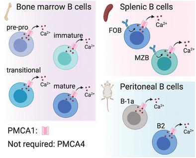
The amplitude and duration of Ca2+ signaling is crucial for B‐cell development and self‐tolerance; however, the mechanisms for terminating Ca2+ signals in B cells have not been determined. In lymphocytes, plasma membrane Ca2+ ATPase (PMCA) isoforms 1 and 4 (PMCA1 and PMCA4, aka ATP2B1 and ATP2B4) are the main candidates for expelling Ca2+ from the cell through the plasma membrane. We report here that Pmca4 (Atp2b4) KO mice had normal B‐cell development, while mice with a conditional KO of Pmca1 (Atp2b1) had greatly reduced numbers of B cells, particularly splenic follicular B cells, marginal zone B cells, and peritoneal B‐1a cells. Mouse and naïve human B cells showed only PMCA1 expression and no PMCA4 by western blot, in contrast to T cells, which did express PMCA4. Calcium handling was normal in Pmca4−/− B cells, but Pmca1 KO B cells had elevated basal levels of Ca2+, elevated levels in ER stores, and reduced Ca2+ clearance. These findings show that the PMCA1 isoform alone is required to ensure normal B‐cell Ca2+ signaling and development, which may have implications for therapeutic targeting of PMCAs and Ca2+ in B cells.
中文翻译:

质膜 Ca2+ ATPase 1 (PMCA1) 但不是 PMCA4 对小鼠的 B 细胞发育和 Ca2+ 稳态至关重要
Ca 2+信号的幅度和持续时间对 B 细胞发育和自我耐受至关重要;然而,终止B 细胞中Ca 2+信号的机制尚未确定。在淋巴细胞中,质膜 Ca 2+ ATPase (PMCA) 亚型 1 和 4(PMCA1 和 PMCA4,又名 ATP2B1 和 ATP2B4)是通过质膜从细胞中排出 Ca 2+的主要候选物。我们在此报告Pmca4 ( Atp2b4 ) KO 小鼠的 B 细胞发育正常,而Pmca1 ( Atp2b1) 的 B 细胞数量大大减少,特别是脾滤泡 B 细胞、边缘区 B 细胞和腹膜 B-1a 细胞。与表达 PMCA4 的 T 细胞相比,小鼠和初始人类 B 细胞通过蛋白质印迹仅显示 PMCA1 表达而未显示 PMCA4。Pmca4 -/- B 细胞的钙处理正常,但Pmca1 KO B 细胞的基础 Ca 2+水平升高,ER 储存水平升高,Ca 2+清除率降低。这些发现表明,仅需要 PMCA1 亚型来确保正常的 B 细胞 Ca 2+信号传导和发育,这可能对B 细胞中 PMCA 和 Ca 2+ 的治疗靶向有影响。
更新日期:2020-10-24
中文翻译:
质膜 Ca2+ ATPase 1 (PMCA1) 但不是 PMCA4 对小鼠的 B 细胞发育和 Ca2+ 稳态至关重要
Ca 2+信号的幅度和持续时间对 B 细胞发育和自我耐受至关重要;然而,终止B 细胞中Ca 2+信号的机制尚未确定。在淋巴细胞中,质膜 Ca 2+ ATPase (PMCA) 亚型 1 和 4(PMCA1 和 PMCA4,又名 ATP2B1 和 ATP2B4)是通过质膜从细胞中排出 Ca 2+的主要候选物。我们在此报告Pmca4 ( Atp2b4 ) KO 小鼠的 B 细胞发育正常,而Pmca1 ( Atp2b1) 的 B 细胞数量大大减少,特别是脾滤泡 B 细胞、边缘区 B 细胞和腹膜 B-1a 细胞。与表达 PMCA4 的 T 细胞相比,小鼠和初始人类 B 细胞通过蛋白质印迹仅显示 PMCA1 表达而未显示 PMCA4。Pmca4 -/- B 细胞的钙处理正常,但Pmca1 KO B 细胞的基础 Ca 2+水平升高,ER 储存水平升高,Ca 2+清除率降低。这些发现表明,仅需要 PMCA1 亚型来确保正常的 B 细胞 Ca 2+信号传导和发育,这可能对B 细胞中 PMCA 和 Ca 2+ 的治疗靶向有影响。


 京公网安备 11010802027423号